Microcirculation: The Liver Acinus
The acinus is the functional microvascular unit of the liver.
It forms around a vertical axis (the portal canal) which consists of a hepatic arteriole,
a portal venule, a bile ductule, as well as lymph vessels and nerves. Blood flow
is vertical to the portal triads and directed radially toward the central veins.
The acinus has three circulatory zones ( Fig.
19-6
),[5]
which reflect the closeness
of hepatocytes to the portal axis or central vein. Zone 1 (periportal zone) cells
are closest to the portal axis (i.e., origin of the sinusoid) and receive blood that
is rich in oxygen and nutrients. Zone 2 (midzonal region) is the arbitrary intermediary
transition zone. Zone 3 (pericentral zone) cells occupy the margin of the acinus,
and receive blood that has exchanged gases and metabolites with cells in zones 1
and 2.
This microvascular architecture creates metabolic diversity among
the three zones. Compartmentalization of metabolic pathways in acinar zones can
promote the efficient use and clearance of metabolic by-products.[12]
[13]
Nitrogen metabolism provides an example.
Zone
1 and zone 2 hepatocytes are the chief producers of ammonia, which is formed when
amino acids are converted to ketoacids. These hepatocytes contain the enzymes of
the urea cycle—a high-capacity, low-affinity cyclical pathway that eliminates
ammonia by incorporating it into urea. If ammonia molecules get through zone 2,
they encounter glutamine synthetase in zone 3. This enzyme is expressed only in
zone 3 and acts to recapture ammonia in the form of glutamine substrate. If the
enzyme were present in zones 1 or 2, ammonia excretion via the urea cycle would be
compromised. However, its presence in zone 3 empowers pericentral hepatocytes to
be important scavengers of ammonia and prevents this toxic substance from reaching
the central circulation.[12]
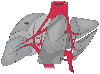 Figure 19-2
Segmental divisions of the liver, with Couinaud's nomenclature.
(Reprinted with permission from Parks RW, Chrysos E, Diamond T: Br J Surg
86:1121, 1999.)
Figure 19-2
Segmental divisions of the liver, with Couinaud's nomenclature.
(Reprinted with permission from Parks RW, Chrysos E, Diamond T: Br J Surg
86:1121, 1999.)
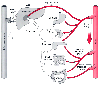 Figure 19-3
The splanchnic circulation. (Redrawn with permission
from Gelman S, Mushlin PS: Catecholamine induced changes in the splanchnic circulation
affecting systemic hemodynamics. Anesthesiology 100:434–439, 2004.)
Figure 19-3
The splanchnic circulation. (Redrawn with permission
from Gelman S, Mushlin PS: Catecholamine induced changes in the splanchnic circulation
affecting systemic hemodynamics. Anesthesiology 100:434–439, 2004.)
Zone 1 hepatocytes have a high proportion of mitochondria and
are the major contributors to oxidative metabolism and glycogen synthesis. In contrast,
zone 3 hepatocytes have large amounts of smooth endoplasmic reticulum, reduced nicotinamide
adenine dinucleotide phosphate (NADPH), and cytochrome P-450 proteins, and are specialized
for anaerobic metabolism and biotransforming xenobiotics. Centrilobular hepatocytes
are therefore the most likely to be injured by toxic intermediates of xenobiotic
metabolism or circulatory disturbances (i.e., ischemia, hypoxia, passive congestion).
Thus, ischemic events often induce decreases of drug metabolism.