Summary of Local Anesthetic Mechanisms
Impulse blockade by local anesthetics may be summarized by the
following chronology[4]
:
- Solutions of local anesthetic are deposited near the nerve. Diffusion
of drug molecules away from this locus is a function of tissue binding, removal by
the circulation, and local hydrolysis of aminoester anesthetics. The net result
is penetration of the nerve sheath by the remaining drug molecules.
- Local anesthetic molecules then permeate the nerve's axon membranes and
reside there and in the axoplasm. The speed and extent of these processes depend
on a particular drug's pKa
and the lipophilicity
of its base and cation species.
- Binding of local anesthetic to sites on voltage-gated Na+
channels
prevents opening of the channels by inhibiting the conformational changes that underlie
channel activation. Local anesthetics bind in the channel's pore and also occlude
the path of Na+
ions.
- During onset and recovery from local anesthesia, impulse blockade is incomplete,
and partially blocked fibers are further inhibited by repetitive stimulation, which
produces an additional, use-dependent binding to Na+
channels.
- One local anesthetic binding site on the Na+
channel may be
sufficient to account for the drug's resting (tonic) and use-dependent (phasic) actions.
Access to this site may potentially involve multiple pathways, but for clinical
local anesthetics, the primary route is the hydrophobic approach from within the
axon membrane.
- The clinically observed rates of onset and recovery from blockade are governed
by the relatively slow diffusion of local anesthetic molecules into and out of the
whole nerve, not by their much faster binding and dissociation to ion channels.
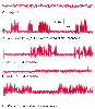 Figure 14-10
Spontaneous ectopic impulses in "abnormal" nerves are
blocked by very low concentrations of local anesthetics. When 10% to 20% of the
Na+
channels close very slowly, as caused here by the addition of a peptide
neurotoxin (ATX), but otherwise an intrinsic property of channels that are upregulated
after nerve injury, spontaneous impulses appear (trace B). Occurring as bursts of
action potentials separated by quiet periods, these impulses appear similar to the
ectopic discharges in a peripheral nerve after injury. Very low concentrations of
lidocaine, equal to plasma levels during intravenous infusions that can reverse neuropathic
pain, strongly suppress (C) and eventually abolish (D) these spontaneous discharges,
an effect that is reversed when lidocaine is removed from the nerve (E). In contrast,
electrically stimulated action potentials are unaffected by such low lidocaine concentrations
(e.g., see Fig 14-8B
).
(From Persaud N, Strichartz G: Micromolar lidocaine selectively blocks propagating
ectopic impulses at a distance from their site of origin. Pain 99:333–340,
2002.)
Figure 14-10
Spontaneous ectopic impulses in "abnormal" nerves are
blocked by very low concentrations of local anesthetics. When 10% to 20% of the
Na+
channels close very slowly, as caused here by the addition of a peptide
neurotoxin (ATX), but otherwise an intrinsic property of channels that are upregulated
after nerve injury, spontaneous impulses appear (trace B). Occurring as bursts of
action potentials separated by quiet periods, these impulses appear similar to the
ectopic discharges in a peripheral nerve after injury. Very low concentrations of
lidocaine, equal to plasma levels during intravenous infusions that can reverse neuropathic
pain, strongly suppress (C) and eventually abolish (D) these spontaneous discharges,
an effect that is reversed when lidocaine is removed from the nerve (E). In contrast,
electrically stimulated action potentials are unaffected by such low lidocaine concentrations
(e.g., see Fig 14-8B
).
(From Persaud N, Strichartz G: Micromolar lidocaine selectively blocks propagating
ectopic impulses at a distance from their site of origin. Pain 99:333–340,
2002.)
A clinically effective block that may last for hours can be achieved
with local anesthetic drugs that dissociate from Na+
channels in a few
seconds.