Electroencephalography
Increasing concentrations of inhaled anesthetics produce a continuum
of electroencephalography (EEG) changes, eventually resulting in burst suppression
and a flat EEG (also see Chapter 31
).
In contrast, a ceiling effect is reached with opioids. Increasing opioid dosage,
once this ceiling has been obtained, does not further affect the EEG.[71]
Problems with lead placement and signal processing need to be resolved before EEG
analysis can be used as a routine monitor of opioid anesthesia depth. It was reported
that emergence from isoflurane but not fentanyl anesthesia is associated with obvious
changes in the overall EEG frequency power spectrum.[72]
Although potency and rate of equilibrium between plasma and brain
are different among opioids, the effects of fentanyl, alfentanil, sufentanil, and
remifentanil are consistent.[73]
Small doses of
fentanyl (200 µg) produce minimal EEG changes, whereas higher doses (30–70
µg/kg) result in high-voltage slow (delta) waves, suggesting a state consistent
with anesthesia. Although transient isolated (usually frontotemporal) sharp wave
activity can be observed after large doses of fentanyl and other opioids, it is not
generalized. Sufentanil produces EEG changes similar to fentanyl ( Fig.
11-6
).[74]
The effects of alfentanil
on the EEG are slightly different from those of fentanyl or sufentanil. Alfentanil
(125 µg/kg) produces less synchronization of the EEG and less change in the
relative EEG power in the delta band.[75]
As an effect site measure, EEG can be employed to assess onset
of drug action and drug potency ratios. It was reported that a lag time between
plasma drug concentration and change in the spectral edge is only 1 min for alfentanil
and 6 min for fentanyl.[74]
The serum concentration
ratio that results in a similar EEG pattern is 75:1 for alfentanil and fentanyl,
[74]
and the lag time is as short for alfentanil
as it is for remifentanil ( Fig. 11-7
).
[76]
This contrasts with the reported dose ratio
in which fentanyl was only three to five times as potent as alfentanil. The serum
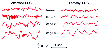 Figure 11-6
Electroencephalography (EEG) stages for fentanyl and
alfentanil. Representative 3-second tracings of EEG recordings from fentanyl and
alfentanil are shown. Awake = mixed α (8–13 Hz) and β (>13 Hz)
activity. Stage 1 = slowing with α spindles. Stage 2 = more slowing, theta
activity present (4–7 Hz). Stage 3 = maximal slowing, δ waves present
(<4 Hz), with high amplitude. (From Scott JC, Ponganis KV, Stanski DR:
EEG quantitation of narcotic effect: The comparative pharmacodynamics of fentanyl
and alfentanil. Anesthesiology 62:234–241, 1985.)
Figure 11-6
Electroencephalography (EEG) stages for fentanyl and
alfentanil. Representative 3-second tracings of EEG recordings from fentanyl and
alfentanil are shown. Awake = mixed α (8–13 Hz) and β (>13 Hz)
activity. Stage 1 = slowing with α spindles. Stage 2 = more slowing, theta
activity present (4–7 Hz). Stage 3 = maximal slowing, δ waves present
(<4 Hz), with high amplitude. (From Scott JC, Ponganis KV, Stanski DR:
EEG quantitation of narcotic effect: The comparative pharmacodynamics of fentanyl
and alfentanil. Anesthesiology 62:234–241, 1985.)
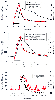 Figure 11-7
Time course of spectral edge and serum opioid concentration.
Fentanyl (top panel) and alfentanil (middle
panel) were infused at 150 µg/minute and 1500 µg/minute, respectively.
Remifentanil (bottom panel) was administered at
3 µg/kg/minute for 10 minutes. The spectral edge changes lag behind the serum
concentration changes in the case of fentanyl, whereas the spectral edge and serum
concentrations are closely parallel in the cases of alfentanil and remifentanil.
(From Scott JC, Ponganis KV, Stanski DR: EEG quantitation of narcotic effect:
The comparative pharmacodynamics of fentanyl and alfentanil. Anesthesiology 62:234–241,
1985; and Egan TD, Minto CF, Hermann DJ, Barr J, Muir KT, Shafer SL: Remifentanil
versus alfentanil: Comparative pharmacokinetics and pharmacodynamics in healthy
adult male volunteers. Anesthesiology 84:821–833, 1996.)
Figure 11-7
Time course of spectral edge and serum opioid concentration.
Fentanyl (top panel) and alfentanil (middle
panel) were infused at 150 µg/minute and 1500 µg/minute, respectively.
Remifentanil (bottom panel) was administered at
3 µg/kg/minute for 10 minutes. The spectral edge changes lag behind the serum
concentration changes in the case of fentanyl, whereas the spectral edge and serum
concentrations are closely parallel in the cases of alfentanil and remifentanil.
(From Scott JC, Ponganis KV, Stanski DR: EEG quantitation of narcotic effect:
The comparative pharmacodynamics of fentanyl and alfentanil. Anesthesiology 62:234–241,
1985; and Egan TD, Minto CF, Hermann DJ, Barr J, Muir KT, Shafer SL: Remifentanil
versus alfentanil: Comparative pharmacokinetics and pharmacodynamics in healthy
adult male volunteers. Anesthesiology 84:821–833, 1996.)
concentration ratio of fentanyl and sufentanil was 12:1 at half-maximal EEG slowing.
[77]
Similar studies suggest that fentanyl and
remifentanil
are 75 and 16 times as potent as alfentanil, respectively.[76]
Potency ratios based on EEG studies are similar to those obtained from studies determining
the plasma drug levels of each opioid necessary to reduce the MAC of isoflurane by
50%. Potency ratios for reduction of the isoflurane MAC for sufentanil:fentanyl:remifentanil:alfentanil
are nearly 1:1/10:1/10:1/100.[62]
[78]
[79]
[80]