RADIATION
During the course of patient management, the anesthesiologist
is almost routinely exposed to both ionizing and nonionizing electromagnetic radiation.
The former is primarily from x-rays and occasionally from radioactive isotopes that
release gamma rays, and the latter is from lasers. Less common is contact with ionizing
radiation from radioactive isotopes that release either alpha or beta particles.
Ionizing radiation has enough energy to create both free radicals and ionized molecules
in tissues by driving electrons completely out of their stable orbitals. If the
radiation exposure is severe enough, tissues may be destroyed or chromosomal changes
may cause malignant growth. Nonionizing radiation may excite electrons to move from
the ground state to higher orbitals in molecules, but the electrons remain in the
molecule. In this case, damage to tissues may result from the heat produced by the
absorbed radiation.
Ionizing Radiation: X-rays
The increasing use of x-rays during the past 2 decades in the
operating room and in remote locations has revolutionized the practices of several
surgical specialties. Fluoroscopy coupled with image intensifiers and video displays
has significantly improved the surgical care of patients by providing immediate information
to the neurosurgeon, orthopedic surgeon, urologist, gastroenterologist, and others.
It may also have decreased operating time by preventing the delays inherent in waiting
for films to be processed. The disadvantage of this increased use of fluoroscopy
is exposure of operating room personnel to ionizing radiation. Because this type
of radiation is undetectable with our normal senses, it occasionally creates undue
fear, called radiation mystique by some. A basic
understanding of its features may reduce some of the unwarranted concerns while permitting
one to keep personal exposure to a minimum. Film badges, though not protective,
provide a means of monitoring exposure.
Exposure is commonly reported in units of rem (roentgen equivalents
man). A rem is essentially a measure of the biologic damage from radiation adjusted
to apply to all tissues.[17]
Estimates of radiation
exposure from natural sources vary, depending on geographic location. The average
in the United States ranges from 80 to 200 millirems (mrem) per year. Natural radiation
comes primarily from cosmic rays (about 40 mrem at sea level, with an increase of
10 mrem/1000 ft), as well as from radioactive compounds found in soil, brick, and
concrete. An isotope of potassium contained in body fluids is an internal source
of beta particles. For most physicians, the additional radiation from occupational
exposure is no greater than that from natural sources. Although the maximum yearly
occupational exposure is mandated to be no more than 5 rem, radiology personnel rarely
absorb more than 10% of this dose. Their greatest source of exposure is fluoroscopy.
[18]
During pregnancy, radiograph personnel are
advised to limit their exposure to a maximum of 500 mrem.
Occupational exposure to radiation comes primarily from x-rays
scattered by both the patient and the surrounding equipment. One chest radiograph
results in about 25 mrem of exposure to the patient; procedures requiring multiple
films occasionally involve more than 1 rem. The amount of radiation generated during
fluoroscopy depends on how long the x-ray beam is on. Although radiograph machines
are designed to minimize stray radiation, some radiation scatters and is absorbed
by personnel who are near the patient. Just as light is reflected from surfaces,
x-rays are reflected from the surfaces on which they impinge ( Fig.
88-1
). This scattering accounts for most occupational exposure. Because
the intensity of scattered radiation is inversely proportional to the square of the
distance from the source, the best protection is physical separation. A distance
of at least 3 feet from the patient is recommended. Six feet of air provides protection
the equivalent of 9 inches of concrete or 2.5 mm of lead.[19]
Although they are uncomfortable, aprons containing the equivalent of 0.25 to 0.5
mm of lead sheet are effective in blocking most scattered radiation, and such devices
are recommended to be worn whenever there is an exposure risk.[20]
Uncovered areas, such as the lens of the eye, still bear the risk of injury. Intraoperative
radiation measurements have shown that exposure is inversely related to the experience
of the surgeon and also that the amount of radiation received by the anesthesiologist
during orthopedic procedures is unmeasurable.[21]
Radiation physicists recommend adhering to the ALARA program:
radiation exposure should be kept as low as reasonably achievable.
The staff of departments of radiology have been extensively schooled in the physics
of radiation as applied both to its diagnostic use and to the safety of occupational
exposure. Attention to their methods of protection will limit exposure to minimal
levels for all. As questions of radiation safety arise, they should be directed
to the health physicists at your hospital.
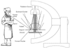 Figure 88-1
Scattering of radiation. Most of the radiation striking
the target and table is reflected upward; some of it passes through the patient and
strikes the detector. Personnel adjacent to the table are struck by scattered radiation.
Figure 88-1
Scattering of radiation. Most of the radiation striking
the target and table is reflected upward; some of it passes through the patient and
strikes the detector. Personnel adjacent to the table are struck by scattered radiation.