PULMONARY RESUSCITATION
For pulmonary resuscitation, the trachea should be intubated immediately,
and positive-pressure ventilation should be initiated at a rate of 30 to 60 breaths/min.
Every fifth breath should be held for 2 to 3 seconds to expand atelectatic lung
and to help remove lung fluid. A PEEP of 1 to 3 mm Hg should be maintained. Care
must be taken not to use excessive pressures to ventilate the lungs of neonates in
the delivery room. Evidence demonstrates that as few as six large breaths at birth
markedly increases the amount of lung injury seen in premature lambs and that the
response to surfactant is significantly diminished in those lambs ventilated with
large breaths.[64]
Excessive inspiratory volumes
are associated with the development of inflammation and chronic lung disease.[8]
The airway pressure should be measured to avoid generating excessive pressures.
This can be done with a pressure manometer or with a pressure transducer.
Tracheal Intubation
The neonate's larynx is located four to six vertebrae more cephalad
than the larynx of adults; consequently, the larynx of the neonate is more anterior
than that of adults. Extension at the head places the larynx in an even more anterior
position and makes tracheal intubation more difficult. The head therefore should
be placed in a neutral or "sniffing" position during bag-and-mask ventilation and
during tracheal intubation. The laryngoscope should be held with the thumb and index
finger and the chin grasped with the ring and middle fingers of the left hand. This
"welds" the head and hand into a single unit and reduces the likelihood of pharyngeal
lacerations occurring if the neonate's head moves. To improve the view of the larynx,
pressure should be applied over the hyoid bone with the small finger of the left
hand ( Fig. 59-7
). This
maneuver moves the larynx posteriorly and exposes the vocal cords. An appropriate-size
endotracheal tube should be inserted and the tip of the tube placed 1 to 2 cm below
the vocal cords, depending on the size of the neonate. The appropriate-size endotracheal
tube is one that permits a small amount of gas to leak from between the endotracheal
tube and trachea when a pressure of 15 to 25 cm H2
O is generated. This
usually means a 2.5-mm (internal diameter) tube for neonates weighing less than 1.5
kg, a 3.0-mm tube for those between 1.5 and 2.5 kg, and a 3.5-mm tube for those weighing
more than 2.5 kg. Tracheal intubation is usually determined by observing the endotracheal
tube pass through the vocal cords, by observing bilateral chest movement with each
mechanical inspiration, and by observing condensation in the endotracheal tube with
exhalation. With auscultation of the chest and epigastrium, the breath sounds should
be much louder over the chest, and the skin color, heart rate, and SaO2
should improve. Correct positioning of the endotracheal tube in the trachea can
be determined by capnography[65]
and SaO2
.
This is a more accurate method of determining placement of the tube in the trachea,
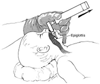 Figure 59-7
Laryngoscopy of the newborn infant. (From Gregory
GA: The Anesthesiologist, Mother, and Newborn. Baltimore, Williams & Wilkins,
1974.)
Figure 59-7
Laryngoscopy of the newborn infant. (From Gregory
GA: The Anesthesiologist, Mother, and Newborn. Baltimore, Williams & Wilkins,
1974.)
and determination of correct tube placement occurs more rapidly with capnography
than with other methods.
The small tidal volumes and the low pulmonary blood flow of some
infants at birth may make it difficult to use capnography effectively. There also
is some CO2
in the expired gas if the endotracheal tube has been inserted
into the esophagus and the neonate was ventilated with a bag and mask initially.