PHYSIOLOGIC CHANGES OF PREGNANCY
Maternal changes in pregnancy occur as a result of hormonal alterations,
mechanical effects of the gravid uterus, increased metabolic and oxygen requirements,
metabolic demands of the fetoplacental unit, and hemodynamic alterations associated
with the placental circulation. Such changes become more significant as pregnancy
progresses, and they have major implications for anesthetic management, especially
in high-risk parturients.
Cardiovascular System
The cardiovascular system adjusts throughout pregnancy to meet
the changes that occur. Hemodynamic and maternal cardiovascular changes in pregnancy
are outlined in Table 58-1
.
Although the physiologic changes in the cardiovascular system appear to begin in
the first trimester, these changes continue into the second and third trimesters,
when cardiac output increases by approximately 40% of nonpregnant values. Cardiac
output increases from the fifth week of pregnancy and reaches its maximum levels
at approximately 32 weeks,
TABLE 58-1 -- Cardiovascular changes in pregnancy
|
Parameter |
Change |
Amount (%) |
|
Heart rate |
Increased |
20–30 |
|
Stroke volume |
Increased |
20–50 |
|
Cardiac output |
Increased |
30–50 |
|
Contractility |
Variable |
±10 |
|
Central venous pressure |
Unchanged |
— |
|
Pulmonary capillary wedge pressure |
Unchanged |
— |
|
Systemic vascular resistance |
Decreased |
20 |
|
Systemic blood pressure |
Slight decrease |
Midtrimester 10–15 mm Hg, then rises |
|
Pulmonary vascular resistance |
Decreased |
30 |
|
Pulmonary artery pressure |
Slight decrease |
— |
|
From Birnbach DJ, Gatt SP, Datta S (eds): Textbook
of Obstetric Anesthesia. New York, Churchill Livingstone, 2000, p 34. |
after which there is only a slight increase until labor, delivery, and the postpartum
period.[5]
Approximately 50% of the increase in
cardiac output has occurred by the eighth week of pregnancy.[6]
Although this increase in cardiac output is due to an increase in both stroke volume
and heart rate, the more important factor is stroke volume, which increases by 20%
to 50% at term from nonpregnant values. Changes in heart rate are extremely difficult
to reliably quantify, but it is thought that the approximately 20% increase in heart
rate is present by the fourth week of pregnancy. Although the normal variability
in heart rate does not change in pregnancy, there does appear to be a reduction in
the sympathetic component.[7]
Tachyarrhythmias
are more common, especially later in pregnancy as a result of both hormonal and autonomic
factors.[8]
Because of the decrease in peripheral vascular resistance, arterial
blood pressure does not change in a normal pregnant patient. Although it was originally
thought that cardiac output decreases during the third trimester, we now know that
this decrease is due to effects of the supine position in the patient at term.[9]
Ueland and colleagues demonstrated that the fall in cardiac output was due to obstruction
of the inferior vena cava by the gravid uterus and did not occur when women were
placed in the lateral position.[10]
A venogram
performed before and after cesarean delivery ( Fig.
58-1
) demonstrates this phenomenon. Despite the increase in blood volume
and cardiac output, parturients at term are susceptible to hypotension, especially
when in the supine position. Up to 10% of pregnant patients at term show signs of
shock when assuming the supine position. This phenomenon has been termed the supine
hypotension syndrome. To compensate, collateral routes of venous return develop,
including the paravertebral veins to the azygos vein. Unlike compression of the
vena cava, compression of the aorta is not associated with maternal symptoms in a
healthy
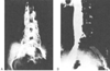 Figure 58-1
A, Venogram in a term
patient in the supine position before cesarean section. Radiopaque dye injected
into the femoral veins fails to reach the inferior vena cava (IVC) but reaches the
paravertebral veins. B, After cesarean section, blood
is seen in the IVC. (From Kerr MG, Scott DB, Samuel E: Studies of the inferior
vena cava in late pregnancy. BMJ 1:532, 1964.)
Figure 58-1
A, Venogram in a term
patient in the supine position before cesarean section. Radiopaque dye injected
into the femoral veins fails to reach the inferior vena cava (IVC) but reaches the
paravertebral veins. B, After cesarean section, blood
is seen in the IVC. (From Kerr MG, Scott DB, Samuel E: Studies of the inferior
vena cava in late pregnancy. BMJ 1:532, 1964.)
parturient, but it may be associated with decreased utero-placental perfusion.[11]
Anesthetics and drugs that cause vasodilation or anesthetic techniques that cause
sympathectomy (neuraxial techniques) may exacerbate aortocaval compression. In the
operating room, a small pillow or "wedge" should be used to provide left uterine
displacement of approximately 15 to 20 degrees. This angle can be increased as necessary
by increasing the wedge or tilting the table.
The changes in blood volume and cardiac output usually have clinical
implications for parturients who have concomitant cardiac disease, but they may also
have an impact on healthy parturients. Many pregnant patients will complain of symptoms
suggestive of cardiovascular disease at term, including shortness of breath, palpitations,
dizziness, edema, and poor exercise tolerance.[12]
Physical examination of the patient may also be abnormal when compared with the
prepregnant state, with auscultation commonly revealing a wide loud split first heart
sound, an S3
sound, and a soft systolic ejection murmur. As illustrated
in Table 58-2
, pregnancy
has numerous effects on cardiac evaluation, including changes in the electrocardiogram,
chest radiograph, and echocardiogram. Although these minor changes occur in healthy
pregnant women at term, symptoms and signs such as chest pain, syncope, severe arrhythmias,
systolic murmur more than grade 3, or diastolic murmur suggest severe disease and
warrant further investigation.[13]
A gradual return
to the prepregnancy blood volume occurs at 6 to 9 weeks postpartum.
 |