PERIOPERATIVE MYOCARDIAL ISCHEMIA
Etiology and Prevention
Ischemic cardiac morbidity is the most common cause of perioperative
(and overall) death in the United States.[26]
Myocardial
ischemia results from an imbalance between myocardial oxygen supply and demand, for
which there are many causes during the perioperative period.[123]
The determinants of myocardial oxygen supply and demand are shown in Figure
52-5
. The most detrimental changes (tachycardia, hypervolemia, and anemia)
are those that simultaneously decrease oxygen supply and increase oxygen demand.
Tachycardia increases myocardial oxygen demand by increasing myocardial work, while
at the
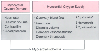 Figure 52-5
Determinants of myocardial oxygen supply and demand that
lead to myocardial ischemia. During the perioperative period, virtually every determinant
is altered by factors such as fluid shifts, blood loss, pain, catecholamines, altered
coagulability, and ventilatory insufficiency. (Adapted from Beattie C, Fleisher
LA: Perioperative myocardial ischemia and infarction. In
Beattie C, Fleisher LA [eds]: International Anesthesiology Clinics. Boston, Little,
Brown, 1992, pp 12–24.)
Figure 52-5
Determinants of myocardial oxygen supply and demand that
lead to myocardial ischemia. During the perioperative period, virtually every determinant
is altered by factors such as fluid shifts, blood loss, pain, catecholamines, altered
coagulability, and ventilatory insufficiency. (Adapted from Beattie C, Fleisher
LA: Perioperative myocardial ischemia and infarction. In
Beattie C, Fleisher LA [eds]: International Anesthesiology Clinics. Boston, Little,
Brown, 1992, pp 12–24.)
same time myocardial oxygen supply is decreased because diastole is shortened and
most coronary blood flow occurs during diastole. Intravascular volume is equally
important. Hypervolemia increases ventricular wall tension and myocardial oxygen
demand. At the same time, coronary perfusion (oxygen supply) is reduced in the distended
ventricle because the increased left ventricular end-diastolic pressure limits coronary
flow. Anemia can also upset both sides of the supply-and-demand equation.[124]
Decreased oxygen content decreases supply, whereas increased heart rate and cardiac
output from anemia increase demand. The prevention and treatment of perioperative
myocardial ischemia require careful control of these and other determinants of myocardial
oxygen supply and demand, as well as other perioperative physiologic changes that
can precipitate ischemia.
When the entire perioperative period is considered, myocardial
ischemia occurs most commonly postoperatively, and less commonly preoperatively and
intraoperatively.[125]
[126]
Postoperative myocardial ischemia is predominantly the ST depression type,[127]
and is significantly associated with MI.[87]
The
low incidence of intraoperative ischemia may be due to anesthetic suppression of
adrenergic tone,[128]
and the minute-to-minute control
of the hemodynamic and other determinants of myocardial oxygen supply and demand.
In the early postoperative period, the patient is transferred from the highly controlled
environment of the operating room to the postanesthetic care unit or intensive care
unit, where the ratio of physician or nurse to patient is reduced, and control of
hemodynamic variables is less complete. Postoperative ischemia often begins early
in the postoperative period, a time associated with pain, tachycardia, hypertension,
sympathetic discharge, and hypercoagulability.[128]
Most postoperative myocardial ischemia is silent (clinically asymptomatic), as a
result of masking by surgical pain or by opioid analgesia,[84]
[129]
[130]
[131]
and is usually associated with an increase in heart rate.[126]
[127]
The peak incidence of ischemia occurs during
the early (days 0 to 3) versus late (days 4 to 7) postoperative period.[126]
A study using continuous 12-lead electrocardiographic monitoring reported 67% of
ischemic events started within 2 hours from the end of surgery and emergence from
anesthesia.[127]
Table 52-8
summarizes eight studies showing the relative incidence of preoperative, intraoperative,
and postoperative myocardial ischemia. The two studies[79]
[132]
with the lowest rates of intraoperative myocardial
ischemia rigorously controlled intraoperative heart rate and blood pressure by protocol.
Norris and associates[132]
continued this "tight"
hemodynamic control into the postoperative period and reported the lowest rate (15%)
of postoperative myocardial ischemia. These studies suggest that good hemodynamic
control perioperatively plays an important role in reducing myocardial ischemia.
Unless a contraindication exists, I administer β-blockers liberally to keep
the heart rate less than 80 beats/min throughout the perioperative period. Although
it is commonly recommended to maintain the blood pressure within 20% of baseline,
I use a more rational approach that better addresses patients with low or high baseline
blood pressure ( Fig. 52-6
).
*Percent
of patients with myocardial ischemia as detected by continuous Holter monitoring
and ST segment analysis.
†Unpublished
data.
Several clinical studies have increased our understanding of the
clinical variables that precipitate myocardial ischemia. There has been a long-standing
debate over whether the rate pressure product (heart rate × mean arterial blood
pressure)[133]
[134]
or the pressure/rate quotient (mean arterial blood pressure divided by heart rate)
[135]
[136]
is most
correlated with ischemic episodes. Work by Buffington[137]
supports the pressure/rate quotient, showing in a canine model that a quotient of
less than 1.0 was associated with ischemia. In simpler terms, hypotension and tachycardia
are a dangerous combination. In patients undergoing CABG
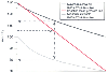 Figure 52-6
Nomogram used to determine minimum and maximum mean blood
pressure limits. The nomogram is used by reading the baseline mean arterial pressure
(MAP) from the left-hand axis horizontally onto the diagonal baseline mean pressure
line. The point of intersection on this line is then extended vertically to intersect
with the minimum and maximum allowable mean pressure curves. A patient with a baseline
MAP of 105 mm Hg, as illustrated, would have an allowable MAP range of 80 to 114
mm Hg. (From Norris EJ, Beattie C, Perler BA, et al: Double-masked randomized
trial comparing alternate combinations of intraoperative anesthesia and postoperative
analgesia in abdominal aortic surgery. Anesthesiology 95:1054–1067, 2001.)
Figure 52-6
Nomogram used to determine minimum and maximum mean blood
pressure limits. The nomogram is used by reading the baseline mean arterial pressure
(MAP) from the left-hand axis horizontally onto the diagonal baseline mean pressure
line. The point of intersection on this line is then extended vertically to intersect
with the minimum and maximum allowable mean pressure curves. A patient with a baseline
MAP of 105 mm Hg, as illustrated, would have an allowable MAP range of 80 to 114
mm Hg. (From Norris EJ, Beattie C, Perler BA, et al: Double-masked randomized
trial comparing alternate combinations of intraoperative anesthesia and postoperative
analgesia in abdominal aortic surgery. Anesthesiology 95:1054–1067, 2001.)
surgery, the pressure/rate quotient was not predictive of ischemia in two different
studies.[138]
[139]
Nonetheless, the importance of heart rate is well recognized as a determinant of
myocardial ischemia in the vascular surgery patient. After vascular surgery, unlike
CABG, patients leave the operating room with the same coronary disease they came
in with, only to experience the stress of the postoperative period. Some vascular
patients exhibit heart rate-related myocardial ischemia at "subtachycardic" heart
rates.[85]
In certain high-risk patients, heart
rates of about 85 beats/min consistently trigger ischemic ST segment changes ( Fig.
52-7
). The primary role of the anesthesiologist is not merely to control
heart rate but also to diagnose and treat the underlying cause of heart rate changes.
The optimal hematocrit value for vascular surgery patients is
unknown. In the mid 1980s, the tendency was to withhold transfusion to avoid the
risk of human immunodeficiency virus and hepatitis infection. In 1988, the National
Institutes of Health indicated that no "threshold" hemoglobin concentration could
be defined for routine transfusion.[140]
The American
College of Physicians then stated that hemoglobin concentrations of greater than
7.0 g/dL are well tolerated in patients without cardiovascular disease.[141]
These guidelines, however, did not include recommendations for patients with CAD
or risk factors for CAD. Although there are no controlled trials, there appears
to be an increased incidence of myocardial ischemia and cardiac morbidity in vascular
surgery patients if hemoglobin concentrations are less than 9.0 g/dL in the early
postoperative period.[142]
[143]
This evidence, along with our understanding of the effects of anemia on myocardial
oxygen supply and demand,[124]
supports the practice
of maintaining hemoglobin concentrations above 9.0 g/dL in the vascular patient,
especially patients at significant risk for ischemic cardiac morbidity.
 |