Discontinuation of Cardiopulmonary Bypass
When separating from CPB, blood volume is assessed by direct visualization
of the heart and monitoring right
atrial or left atrial filling pressures. When filling pressures are adequate, the
patient fully warmed, acid-base status normalized, heart rate adequate, and sinus
rhythm achieved, the venous drainage is stopped and the patient can be weaned from
bypass. The arterial cannula is left in place so that a slow infusion of residual
pump blood can be used to optimize filling pressures. Myocardial function is assessed
by direct cardiac visualization and by a transthoracic left or right atrial catheter,
by a percutaneous internal jugular catheter, or by the use of intraoperative echocardiography.
Pulse oximetry can also be used to assess the adequacy of cardiac output.[154]
Low systemic arterial saturation or the inability of the oximeter probe to register
a pulse may be a sign of very low output and high systemic resistance.[155]
After the repair of complex congenital heart defects, the anesthesiologist
and surgeon may have difficulty separating patients from CPB. Under these circumstances,
a diagnosis must be made and includes (1) an inadequate surgical result with a residual
defect requiring repair, (2) pulmonary artery hypertension, and (3) right or left
ventricular dysfunction.
Two general approaches are customarily used, either independently
or in conjunction. An intraoperative "cardiac catheterization" can be performed
to assess isolated pressure measurements from the various great vessels and chambers
of the heart (i.e., catheter pullback measurements or direct needle puncture to evaluate
residual pressure gradients across repaired valves, sites of stenosis and conduits,
and oxygen saturation data to examine for residual shunts).[156]
Alternatively, echo-Doppler may be used to provide an intraoperative image of structural
or functional abnormalities to assist in the evaluation of the postoperative cardiac
repair.[16]
[157]
If structural abnormalities are found, the patient can be placed back on CPB, and
residual
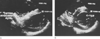 Figure 51-12
A, Two-dimensional echocardiogram
in the short-axis view across the ventricles demonstrating the presence of intramyocardial
air (arrow) in the ventricular septum and right ventricular
wall. The intramyocardial air appears as a dense, "snowy" echogenic area. Note
the associated wall motion abnormality appearing as flattening of the ventricular
septum. B, The patient was treated with phenylephrine,
increasing systemic and coronary perfusion pressure, resulting in clearance of the
air and the echogenic density and restoration of normal left ventricular (LV) wall
motion and configuration.
Figure 51-12
A, Two-dimensional echocardiogram
in the short-axis view across the ventricles demonstrating the presence of intramyocardial
air (arrow) in the ventricular septum and right ventricular
wall. The intramyocardial air appears as a dense, "snowy" echogenic area. Note
the associated wall motion abnormality appearing as flattening of the ventricular
septum. B, The patient was treated with phenylephrine,
increasing systemic and coronary perfusion pressure, resulting in clearance of the
air and the echogenic density and restoration of normal left ventricular (LV) wall
motion and configuration.
defects can be repaired before the patient leaves the operating room. Leaving the
operating room with a significant residual structural defect adversely affects survival
and increases patient morbidity (see Fig.
51-5
).[16]
[157]
Echo-Doppler can rapidly identify right and left ventricular dysfunction and suggest
the presence of pulmonary artery hypertension. In addition, echo-Doppler can identify
regional wall motion abnormalities due to ischemia or intramyocardial air that will
direct specific pharmacologic therapy and provide a means of assessing the results
of these interventions ( Fig. 51-12
).
[158]