|
|
|
|
|
|
|
|
|
|
|
|
|
|
|
Mitral stenosis (MS) is almost always rheumatic in origin. Thus, though still a common indication for mitral valve replacement, its overall incidence in developed countries continues to decline with improved socioeconomic conditions. MS exhibits a 2:1 female preponderance. Infrequent causes include congenital defects (seen in infancy and childhood), malignant carcinoid (although right heart lesions are its usual cardiac manifestation), rheumatoid arthritis, and prolapse of left atrial myxomas into the mitral valve opening.
Although rheumatic fever causes pancarditis, the long-term sequelae are confined to the endocardium (including the valves). Pure MS occurs in 25% of patients who develop long-term rheumatic sequelae. The remaining patients have either combined lesions in one valve (e.g., combined stenosis and regurgitation occur in 40% of patients) or combined valvular disease (aortic plus mitral disease). Rheumatic-induced chronic aortic valve disease never occurs in isolation; concurrent mitral valve disease is also present. Although the interval between rheumatic fever and the development of MS is at least 2 years, patients are usually asymptomatic for 2 decades and symptoms generally develop in the 3rd and 4th decades of life. Progression is much more rapid in underdeveloped countries. It is unclear whether the chronic valvular sequelae of rheumatic fever reflect ongoing smoldering rheumatic fever or the consequences of turbulent blood flow patterns induced by the initial rheumatic process in a manner analogous to the development of aortic stenosis in a patient with a bicuspid aortic valve. In patients with chronic
 Figure 50-22
Hemodynamics of aortic regurgitation. A,
Normal conditions. B, The hemodynamic changes that
occur in severe acute aortic regurgitation. Although total stroke volume is increased,
forward stroke volume is reduced. Left ventricular end-diastolic pressure rises
dramatically. C, Hemodynamic changes occurring in
chronic compensated aortic regurgitation are shown. Eccentric hypertrophy produces
increased end-diastolic volume, which permits an increase in total as well as forward
stroke volume. The volume overload is accommodated, and left ventricular filling
pressure is normalized. Ventricular emptying and end-systolic volume remain normal.
D, In chronic decompensated aortic regurgitation,
impaired left ventricular emptying produces an increase in end-systolic volume and
a fall in ejection fraction, total stroke volume, and forward stroke volume. Further
cardiac dilatation occurs along with re-elevation of left ventricular filling pressure.
E, Immediately after valve replacement, preload estimated
by end-diastolic volume decreases, as does filling pressure. End-systolic volume
is also decreased, but to a lesser extent. The result is an initial fall in ejection
fraction. Despite these changes, elimination of regurgitation leads to an increase
in forward stroke volume. AoP, aortic pressure; EDV, end-diastolic volume; EF, ejection
fraction; ESV, end-systolic volume; LVEDP, left ventricular end-diastolic pressure;
RF, regurgitant fraction. (From Carabello BA: Aortic regurgitation: Hemodynamic
determinants of prognosis. In Cohn LH, DiSesa VJ
[eds]: Aortic Regurgitation: Medical and Surgical Management. New York, Marcel
Dekker, 1986.)
Figure 50-22
Hemodynamics of aortic regurgitation. A,
Normal conditions. B, The hemodynamic changes that
occur in severe acute aortic regurgitation. Although total stroke volume is increased,
forward stroke volume is reduced. Left ventricular end-diastolic pressure rises
dramatically. C, Hemodynamic changes occurring in
chronic compensated aortic regurgitation are shown. Eccentric hypertrophy produces
increased end-diastolic volume, which permits an increase in total as well as forward
stroke volume. The volume overload is accommodated, and left ventricular filling
pressure is normalized. Ventricular emptying and end-systolic volume remain normal.
D, In chronic decompensated aortic regurgitation,
impaired left ventricular emptying produces an increase in end-systolic volume and
a fall in ejection fraction, total stroke volume, and forward stroke volume. Further
cardiac dilatation occurs along with re-elevation of left ventricular filling pressure.
E, Immediately after valve replacement, preload estimated
by end-diastolic volume decreases, as does filling pressure. End-systolic volume
is also decreased, but to a lesser extent. The result is an initial fall in ejection
fraction. Despite these changes, elimination of regurgitation leads to an increase
in forward stroke volume. AoP, aortic pressure; EDV, end-diastolic volume; EF, ejection
fraction; ESV, end-systolic volume; LVEDP, left ventricular end-diastolic pressure;
RF, regurgitant fraction. (From Carabello BA: Aortic regurgitation: Hemodynamic
determinants of prognosis. In Cohn LH, DiSesa VJ
[eds]: Aortic Regurgitation: Medical and Surgical Management. New York, Marcel
Dekker, 1986.)
The essential characteristics of a “normal” mitral valve are
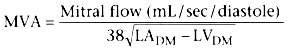
The Gorlin formula[130] is used to calculate mitral valve area and dictates, as inferred earlier, that flow and the
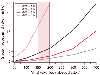 Figure 50-23
The gradient across mitral valves of various sizes is
plotted according to mitral valve flow. Note that when mitral valve flow is in the
normal range (150 to 200 mL/sec of diastole), the gradient across a mitral valve
area (MVA) of 2 cm2
is 4 to 6 mm Hg. With an MVA of 1 cm2
,
the gradient with normal mitral valve flow is 18 to 28 mm Hg. (Redrawn from
Dalen JE, Alpert JS [eds]: Valvular Heart Disease, 2nd ed. Boston, Little, Brown,
1987.)
Figure 50-23
The gradient across mitral valves of various sizes is
plotted according to mitral valve flow. Note that when mitral valve flow is in the
normal range (150 to 200 mL/sec of diastole), the gradient across a mitral valve
area (MVA) of 2 cm2
is 4 to 6 mm Hg. With an MVA of 1 cm2
,
the gradient with normal mitral valve flow is 18 to 28 mm Hg. (Redrawn from
Dalen JE, Alpert JS [eds]: Valvular Heart Disease, 2nd ed. Boston, Little, Brown,
1987.)
Left ventricular end-diastolic pressure and volume are usually normal in patients with pure MS, although left ventricular end-diastolic volume is decreased in a minority of patients. Indices of overall systolic function indicate that systolic function is compromised in up to 25% of patients. Moreover, regional wall motion abnormalities are common. These changes may reflect the underlying rheumatic process or concomitant coronary artery disease and may explain why a subpopulation of patients with MS do poorly after mitral valve replacement.
Patients with MS may have cardiac symptoms or noncardiac symptoms secondary to thromboembolic phenomena. The dominant cardiac symptoms reflect the underlying pathophysiology, specifically, the ability to maintain normal cardiac output and the status of the pulmonary vasculature. Patients with moderate stenosis (mitral valve area of 1 to 1.5 cm2 ) may not necessarily have significant secondary pulmonary vasculature changes, but experience profound dyspnea secondary to pulmonary congestion if the valvular gradient increases acutely (e.g., new-onset atrial fibrillation, increased cardiac output with exercise). Conversely, patients with severe stenosis (mitral valve area less than 1 cm2 ) are likely to have significant secondary pulmonary vascular changes and an inability to generate an adequate cardiac output and thus complain primarily of fatigue and weakness. Patients with MS are always at risk of infective endocarditis and may have hemoptysis or compression of local structures secondary to left atrial expansion (e.g., recurrent laryngeal nerve and esophageal compression leading to dysphagia).
Patients with asymptomatic MS are managed medically, whereas those with mild symptoms should have their management customized to their specific medical and domestic circumstances. Patients with symptomatic critical stenosis and those with thromboembolic phenomena are managed surgically. Surgical options include closed balloon commissurotomy, closed and open surgical commissurotomy, and mitral valve replacement. All commissurotomy procedures are associated with mitral regurgitation and thromboembolism, although this intervention may be appropriate in selected patients, such as those with little calcification, minimal mitral regurgitation, or subvalvular disease involvement.[146]
Successful intraoperative management of patients with MS requires not only knowledge of what constitutes a normal/abnormal mitral valve area but also an understanding of (1) the implications of mitral annular and mitral valve calcification; (2) the possibility of concurrent non-mitral valve disease; (3) the fundamental interaction between flow, diastolic time, and the transvalvular mitral valve gradient, plus the implications that will ensue proximally (e.g., left atrial pressure, pulmonary artery pressure, pulmonary congestion), and distally (cardiac output); (4) the influence of rheumatic disease on left ventricular function; (5) the implications of visualizing the left atrial appendage and confirming the presence or absence of thrombus; (6) the reversibility of pulmonary vascular
|
|
|
|
|
|
|
|
|
|
|
|
|