Physiology of Spontaneous Ventilation with an Open
Chest
Mediastinal Shift
An examination of the physiology of the open chest during spontaneous
ventilation reveals why controlled positive-pressure ventilation is the only practical
way to provide adequate gas exchange during thoracotomy. In a spontaneously breathing,
closed-chest patient in the LDP, gravity causes the pleural pressure in the dependent
hemithorax to be less negative than in the nondependent hemithorax,
but there is still negative pressure in each hemithorax on each side of the mediastinum.
In addition, the weight of the mediastinum causes some compression of the lower
lung, thereby contributing to the pleural pressure gradient. With the nondependent
hemithorax open, atmospheric pressure in that cavity exceeds the negative pleural
pressure in the dependent hemithorax; this imbalance in pressure on the two sides
of the mediastinum causes further downward displacement of the mediastinum into the
dependent thorax. During inspiration, the caudad movement of the dependent lung
diaphragm increases the negative pressure in the dependent lung and causes a still
further displacement of the mediastinum into the dependent hemithorax. During expiration,
as the dependent lung diaphragm moves cephalad, the pressure in the dependent hemithorax
becomes relatively positive, and the mediastinum is pushed upward out of the dependent
hemithorax ( Fig. 49-8
).
Thus, the tidal volume in the dependent lung is decreased by an amount equal to
the inspiratory displacement caused by mediastinal movement. This phenomenon is
called mediastinal shift and is one mechanism that results in impaired ventilation
in an open-chest, spontaneously breathing patient in the LDP. The mediastinal shift
can also cause circulatory changes (decreased venous return) and reflexes (sympathetic
activation) that result in a clinical picture similar to shock: the patient is hypotensive,
pale, and
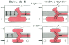 Figure 49-8
Schematic representation of mediastinal shift and paradoxical
respiration in a spontaneously ventilating patient with an open chest who is placed
in the lateral decubitus position. The open chest is always exposed to atmospheric
pressure (+). During inspiration, negative pressure (-) in the intact hemithorax
causes the mediastinum to move downward (mediastinal shift). In addition, during
inspiration, movement of gas from the nondependent lung in the open hemithorax into
the dependent lung in the closed hemithorax and movement of air from the environment
into the open hemithorax cause the lung in the open hemithorax to collapse (paradoxical
respiration). During expiration, relative positive (+) pressure in the closed hemithorax
causes the mediastinum to move upward (mediastinal shift). In addition, during expiration,
gas moves from the dependent lung to the nondependent lung and from the open hemithorax
to the environment; consequently, the nondependent lung expands during expiration
(paradoxical respiration). (From Benumof JL: Anesthesia for Thoracic Surgery.
Philadelphia, WB Saunders, 1987.)
Figure 49-8
Schematic representation of mediastinal shift and paradoxical
respiration in a spontaneously ventilating patient with an open chest who is placed
in the lateral decubitus position. The open chest is always exposed to atmospheric
pressure (+). During inspiration, negative pressure (-) in the intact hemithorax
causes the mediastinum to move downward (mediastinal shift). In addition, during
inspiration, movement of gas from the nondependent lung in the open hemithorax into
the dependent lung in the closed hemithorax and movement of air from the environment
into the open hemithorax cause the lung in the open hemithorax to collapse (paradoxical
respiration). During expiration, relative positive (+) pressure in the closed hemithorax
causes the mediastinum to move upward (mediastinal shift). In addition, during expiration,
gas moves from the dependent lung to the nondependent lung and from the open hemithorax
to the environment; consequently, the nondependent lung expands during expiration
(paradoxical respiration). (From Benumof JL: Anesthesia for Thoracic Surgery.
Philadelphia, WB Saunders, 1987.)
cold, with dilated pupils. Local anesthetic infiltration of the pulmonary plexus
at the hilum and the vagus nerve can diminish these reflexes. More practically,
controlled positive-pressure ventilation abolishes these ventilatory and circulatory
changes associated with mediastinal shift.
Paradoxical Respiration
When the pleural cavity is exposed to atmospheric pressure, the
lung is no longer held open by negative intrapleural pressure, and it tends to collapse
because of unopposed elastic recoil.[225]
Thus,
the lung in an open chest is at least partially collapsed. It has long been observed
during spontaneous ventilation with an open hemithorax that lung collapse is accentuated
during inspiration and, conversely, the lung expands during expiration. This reversal
of lung movement during respiration with an open chest has been termed paradoxical
respiration. The mechanism of paradoxical respiration is similar to that of mediastinal
shift. During inspiration, the descent of the diaphragm on the side of the open
hemithorax causes air from the environment to enter the pleural cavity on that side
through the thoracotomy opening and fill the space around the exposed lung. The
descent of the hemidiaphragm on the closed-chest side causes gas to enter the closed-chest
lung in the normal manner. However, gas also enters the closed-chest lung (which
has a relatively negative pressure) from the open-chest lung (which remains at atmospheric
pressure), which results in a further reduction in the size of the open-chest lung
during inspiration. During expiration the reverse occurs, with the collapsed, open-chest
lung filling from the intact lung and air moving back out of the exposed hemithorax
through the thoracotomy incision. The phenomenon of paradoxical respiration is illustrated
in Figure 49-8
. Paradoxical
breathing is increased by a large thoracotomy and by increased airway resistance
in the intact lung. Paradoxical respiration may be prevented either by manual collapse
of the open-chest lung or, more commonly, by controlled positive-pressure ventilation.
 |