Axillary Approaches to the Brachial Plexus
Axillary blocks are performed with the child placed in the supine
position.[201]
The relevant arm is supinated and
abducted 90 degrees from the body. The landmarks are the pectoralis major muscle,
the coracobrachialis muscle, and the axillary or brachial arteries. Several sites
of puncture have been described, but because of loose fascial attachments, they do
not influence the distribution of
anesthesia in children, even in regard to the musculocutaneous nerve, the block of
which depends on the site of emergence from the perineural space and not on the site
or the technique of puncture. The most usual puncture site is at the upper border
of the axillary artery, as high as possible in the axilla. The needle is inserted
through the skin at a 45-degree angle, pointing toward the midpoint of the clavicle,
until it crosses the perineurovascular sheath with a characteristic click. At this
stage, muscle twitches are elicited in the median or the radial nerve (rarely, the
ulnar nerve), and the local anesthetic can be injected. Whatever the nerve first
identified, contrary to what happens in most adults, complete blockade of these three
nerves is obtained, and there is no advantage in trying to locate each nerve and
in performing multiple injections.[210]
The main limitation of this approach is related to the musculocutaneous
nerve, which remains unchanged in 50% of procedures for anatomic reasons. To overcome
this limitation, I recommend using a transcoracobrachialis approach.[201]
The site of puncture lies at the crossing of the coracobrachialis muscle with the
lower border of the pectoralis major muscle ( Fig.
45-5
). The needle is introduced perpendicular to the skin, moved through
the upper and lateral part of the coracobrachialis muscle (within which runs the
musculocutaneous muscle), and advanced toward the humerus, just above the axillary
artery, which is firmly held by finger compression. The musculocutaneous muscle
is stimulated first (i.e., flexion of the forearm), and the needle is moved forward
until a click is perceived and twitches are elicited in muscles supplied by the median,
radial, or rarely, the ulnar nerve. The local anesthetic is then injected, and while
withdrawing the needle, a small volume (0.5 to 1 mL) of
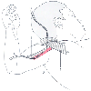 Figure 45-5
Axillary approaches to the brachial plexus: classic
approach (A) and transcoracobrachialis approach (B), indicating the pectoralis major
muscle (1), axillary artery (2), and coracobrachialis muscle (3).
Figure 45-5
Axillary approaches to the brachial plexus: classic
approach (A) and transcoracobrachialis approach (B), indicating the pectoralis major
muscle (1), axillary artery (2), and coracobrachialis muscle (3).
local anesthetic is injected within the substance of the coracobrachialis muscle
to ensure blockade of the musculocutaneous nerve.
Several variants of axillary approaches have been published, but
they are of little interest or are even detrimental in children (especially transaxillary
artery approaches). Some skilled anesthesiologists still use a cannula technique
with excellent results.[211]
As for all block procedures,
performing the block before surgery rather than at the end of the operation improves
the quality of blockade.[212]
Commonly used local
anesthetics are displayed in Table
45-5
. Levobupivacaine, which is still being evaluated in children, seems
promising based on adult data.[213]
Ropivacaine
is commonly used, even though few data have been published in the pediatric literature.
Sound selection of additives, including clonidine,[214]
can improve the quality and duration of blockade, whereas opioids do not offer any
advantage.[215]
When long-lasting pain relief is
mandatory, placement of a catheter is recommended. The technique of inserting an
axillary catheter is easy, provided an adequate device is used, but dressing and
immobilization are rather difficult, especially with classic approaches (easier with
the transcoracobrachialis approach). A coracoid approach may be preferred in this
case, even though the technique is not as easy and safe as the axillary approach.
Recommended volumes of anesthetic solution for single-shot procedures
are shown in Table 45-11
.
Common rates of continuous infusions of local anesthetics used in my institution
are displayed in Table 45-12
.
Regardless of the technique used, axillary blocks are virtually free of complications.
Accidental arterial puncture is the most undesirable occurrence, which may occasionally
result in transient vascular insufficiency or a compressive hematoma. Pneumothorax
has been observed after very inappropriate insertion routes, but it is unusual.