|
|
|
|
|
|
|
|
|
|
|
|
|
|
|
Anesthesia facemasks of rubber or plastic are employed to administer oxygen and anesthetic gases and to ventilate the nonintubated patient. Masks come in a large variety of shapes, but the anatomic mask is most commonly used in adults. Adult masks come in small, medium, and large sizes (nos. 3, 4, and 5). Most adults can be ventilated with a small or medium mask, but occasionally, the patient with a long or wide face or large nose requires a large mask. Children's masks come in newborn, infant, and children sizes. In addition to the anatomic mask, the relatively flat Rendell-Baker-Soucek mask is often used because it conforms to the child's flatter face and has minimal dead space. Transparent masks are being used more often for adults and children. They are less frightening than a black, opaque mask, and the patient can be better observed for cyanosis and vomiting.
The mask is held with one hand, as shown in Figure 42-4 . The fingers should be kept on the bone rather than soft tissues because the latter position can cause discomfort in the awake patient and can cause airway obstruction if such pressure sufficiently raises the base of the tongue. Ventilation with a mask requires a tight fit that involves downward displacement of the mask with the thumb and first finger and upward displacement of the mandible with the other three fingers. Mandibular displacement along with upper cervical extension and chin lift all tend to pull the tongue and soft tissues up off the posterior pharyngeal wall and relieve the upper airway obstruction that occurs in the anesthetized or unconscious patient ( Fig. 42-5 ). This may require holding the mask with two hands and vigorously pulling the mandible upward ("jaw thrust") ( Fig. 42-6 ). A two-handed mask grip requires an assistant to provide manual ventilation. If no such help is available, the anesthesia ventilator can be used to supply positive-pressure breaths. If necessary, the anesthetist's chin can exert downward pressure on the elbow connector to achieve a tight mask fit. Alternatively, the second person can provide jaw thrust while the first seals the mask and ventilates by anesthesia bag.
Mask ventilation may be extremely difficult for patients with problems such as obesity, tumors, infections, and inflammatory disorders. The pediatric patient usually presents less of a problem than the adult because
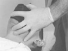 Figure 42-4
Technique for holding the mask with one hand. An effort
should be made to avoid excessive pressure on the soft tissues of the neck.
Figure 42-4
Technique for holding the mask with one hand. An effort
should be made to avoid excessive pressure on the soft tissues of the neck.
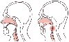 Figure 42-5
A, The normal airway.
The tongue and other soft tissues are forward, allowing an unobstructed air passage.
B, Obstructed airway. The tongue and epiglottis
fall back to the posterior pharyngeal wall, occluding the airway. (Adapted
from Dorsch JA, Dorsch SE: Understanding Anesthesia Equipment, 4th ed. Baltimore,
Williams & Wilkins, 1999.)
Figure 42-5
A, The normal airway.
The tongue and other soft tissues are forward, allowing an unobstructed air passage.
B, Obstructed airway. The tongue and epiglottis
fall back to the posterior pharyngeal wall, occluding the airway. (Adapted
from Dorsch JA, Dorsch SE: Understanding Anesthesia Equipment, 4th ed. Baltimore,
Williams & Wilkins, 1999.)
When airway integrity cannot be maintained with manipulation of the mask, mandible, or neck, a mechanical airway may restore airway patency. Oral and nasal
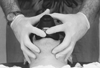 Figure 42-6
Technique for holding the mask with two hands.
Figure 42-6
Technique for holding the mask with two hands.
 Figure 42-7
Oropharyngeal airway in place. The airway follows the
curvature of the tongue, pulling it and the epiglottis away from the posterior pharyngeal
wall and providing a channel for air passage. (Adapted from Dorsch JA, Dorsch
SE: Understanding Anesthesia Equipment, 4th ed. Baltimore, Williams & Wilkins,
1999.)
Figure 42-7
Oropharyngeal airway in place. The airway follows the
curvature of the tongue, pulling it and the epiglottis away from the posterior pharyngeal
wall and providing a channel for air passage. (Adapted from Dorsch JA, Dorsch
SE: Understanding Anesthesia Equipment, 4th ed. Baltimore, Williams & Wilkins,
1999.)
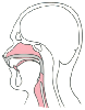 Figure 42-8
The nasopharyngeal airway in place. The airway passes
through the nose and ends at a point just above the epiglottis. (Adapted
from Dorsch JA, Dorsch SE: Understanding Anesthesia Equipment, 4th ed. Baltimore,
Williams & Wilkins, 1999.)
Figure 42-8
The nasopharyngeal airway in place. The airway passes
through the nose and ends at a point just above the epiglottis. (Adapted
from Dorsch JA, Dorsch SE: Understanding Anesthesia Equipment, 4th ed. Baltimore,
Williams & Wilkins, 1999.)
Soft nasal airways are useful in patients who are not deeply anesthetized because such airways tend to provoke less airway stimulation. Relative contraindications to such airways include coagulopathy, basilar skull fracture, and nasal infections or deformities. If possible, vasoconstriction with phenylephrine nose drops (and topical anesthesia with lidocaine if the patient is awake) should precede any nasal instrumentation. However, in the acute situation, the lubricating qualities of lidocaine ointment may suffice. The tip of the airway should be inserted perpendicularly to the face and not upward toward the cribriform plate. It has been suggested that the length of the airway should be roughly the distance from the tip of the nose to the meatus of the ear. Adult nasal airways are measured in French numbers; values are given for the outer diameter and therefore reflect circumferences that range in size from 28 to 30 and 32 to 34. If the nasal airway does not have a sizable flange at the nasal end, the placement of a safety pin (taped to the face) in the airway tip can prevent loss of the nasal airway into the patient's lower airway. Adequately large oral and nasal airways should be employed together if mask ventilation cannot be otherwise accomplished.
|
|
|
|
|
|
|
|
|
|
|
|
|