Acceleromyography
The technique of AMG is based on Newton's second law: force equals
mass times acceleration.[64]
[65]
If mass is constant, acceleration is directly proportional to force. Accordingly,
after nerve stimulation, one can measure not only the evoked force but also the acceleration
of the thumb.
AMG uses a piezoelectric ceramic wafer with electrodes on both
sides. Exposure of the electrode to a force generates an electrical voltage proportional
to the acceleration of the thumb in response to nerve stimulation. When the accelerometer
is fixed to the thumb and the ulnar nerve is stimulated, an electrical signal is
produced whenever the thumb moves. This signal is then analyzed in a specially designed
analyzer[65]
or perhaps displayed on a recording
system. At least one detached monitor based on measurement of acceleration is commercially
available: the TOF-Watch (Organon Teknika, Boxtel, Holland) ( Fig.
39-14
).
AMG is a simple method of analyzing neuromuscular function, both
in the operating room and in the intensive care unit.[66]
[67]
However, although a good correlation exists
between the TOF ratio measured by this method and TOF ratio measured with a force-displacement
transducer or using EMG,[64]
[65]
[68]
measurements made with an AMG are not directly
comparable with results obtained using the other two methods.[69]
[70]
[71]
[72]
[73]
[74]
[75]
When an AMG is used, as originally suggested, with a free-moving thumb,[64]
wide limits of agreements in twitch height (T1) and TOF ratio and differences in
the onset and recovery course of blockade between AMG and MMG have been found. Also,
the AMG control TOF ratio is consistently higher than when measured using a force-displacement
transducer. In accordance with this, one study has indicated that when using AMG,
the threshold TOF ratio for sufficient postoperative neuromuscular recovery is 1.0,
rather than 0.9 as when measured with MMG or EMG.[76]
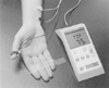 Figure 39-14
TOF-Watch (Organon Ltd., Dublin, Ireland). This neuromuscular
transmission monitor is based on measurement of acceleration using a piezoelectric
transducer.[64]
[65]
Note the transducer fastened to the thumb and the stimulating electrodes. On the
display of the TOF-Watch, the TOF ratio is given in percentage.
Figure 39-14
TOF-Watch (Organon Ltd., Dublin, Ireland). This neuromuscular
transmission monitor is based on measurement of acceleration using a piezoelectric
transducer.[64]
[65]
Note the transducer fastened to the thumb and the stimulating electrodes. On the
display of the TOF-Watch, the TOF ratio is given in percentage.
One reason for the wide limits of agreement between AMG and MMG
is probably and paradoxically connected with one of the originally claimed advantages
of the method, that fixation of the hand could be reduced to a minimum as long as
the thumb could move freely.[64]
However, in daily
clinical practice it is often not possible to ensure that the thumb can move freely,
and that the position of the hand does not change during a surgical procedure. The
evoked response may therefore vary considerably. Several solutions have been proposed,
and ongoing clinical research indicates that the use of an elastic preload to the
thumb may improve the agreement between results obtained using AMG and MMG ( Fig.
39-15
). In spite of the above reservations, it is my opinion that AMG
at the thumb is a valuable clinical tool that used with intelligence may eliminate
the problem of postoperative residual neuromuscular block.[77]
[78]
When the thumb is not available for monitoring during surgery,
some clinicians prefer to monitor the AMG response of the orbicularis oculi or the
corrugator supercilii in response to facial nerve stimulation. However, it is important
to realize that neuromuscular monitoring with AMG from both these sites are subject
to a large uncertainty as a measure of paralysis, and it cannot therefore be recommended
for routine monitoring. It only provides a rough estimate of the degree of block
of the peripheral muscles.[39]
[79]
[80]