Visual Evoked Potentials
VEPs are recorded after monocular stimulation with recording electrodes
over the occipital, parietal, and central scalp.[107]
Pattern reversal of a checkerboard pattern with constant luminance is the preferred
stimulus because the generated evoked potentials have a narrower range of normal
variation and are more sensitive to conduction defects. However, it is not possible
to deliver this type of stimulation intraoperatively to the anesthetized patient.
Instead, flash stimulation of the retina through closed eyelids is provided using
light-emitting diodes embedded in soft plastic goggles. The flash rate is 1 to 3
Hz, with a duration of 3 to 5 msec. The major evoked response peak latencies are
between 90 and 200 msec ( Fig. 38-12
),
and signals are large enough that prolonged averaging is not needed, and frequently,
as few as 20 responses may be averaged.[57]
[79]
[80]
[105]
For
some
procedures, such as operations on the anterior cranial fossa, the goggles interfere
with approach to the operative field. Light-emitting diodes embedded within a plastic
contact lens have been used in these situations.[77]
VEPs are cortical SERs that vary with the type of stimulus, part of the retina stimulated,
degree of pupil dilation, and patient's attention level.[57]
Because some of these factors change commonly and even constantly during the course
of every anesthetic, VEPs would be expected to be highly variable during surgery
even when no surgical trespass on the visual system occurs.
Surgical Procedures Monitored with Visual Evoked Potentials
Intraoperative VEP monitoring has been advocated for procedures
placing the visual system at risk, especially for those in the area of the optic
chiasm. Procedures in which
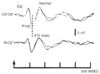 Figure 38-12
Pattern shift of visual evoked potentials. (Adapted
from Chicappa KH, Ropper AH: Evoked potentials in clinical medicine. N Engl J Med
306:1140, 1982.)
Figure 38-12
Pattern shift of visual evoked potentials. (Adapted
from Chicappa KH, Ropper AH: Evoked potentials in clinical medicine. N Engl J Med
306:1140, 1982.)
VEP monitoring has been used include resection of pituitary tumors, craniopharyngioma,
optic glioma, orbital pseudotumor, occipital arteriovenous malformation, meningioma
impinging on the optic chiasm, and chondrosarcoma of the sphenoid wing; drainage
of pituitary abscess; clipping of internal carotid artery and basilar artery aneurysms;
surgical correction of cerebrospinal fluid rhinorrhea; and treatment of orbital fracture.
[77]
[79]
[80]
Changes in evoked potential latency and amplitude were used to guide operative manipulations
or to indicate adequate systemic blood pressure in patients in whom induced hypotension
was being used.[57]
[80]
Intraoperative recordings can be recorded in 88% to 100% of patients.[77]
[79]
[80]
However,
intraoperative variability unrelated to changes in neurologic function may be as
high as 68% to 81%.[79]
[80]
In one large series, there was a relatively high incidence of false-positive and
false-negative results. Thirteen percent of patients with intraoperative loss of
VEPs had unchanged vision postoperatively, and 7% had intact VEPs with significant
visual defects.[80]
VEPs are sensitive to a number of factors that cannot be controlled
intraoperatively. Stimulus delivered to the retina is difficult to control because
the flash must pass through the closed eyelid, and intraoperative changes in pupillary
size and direction are common. Improved systems to deliver the stimulus intraoperatively
need to be developed.[80]
In the opinion of the
investigator of one of the largest series of patients studied with intraoperative
VEP monitoring using current techniques, VEPs cannot be reliably interpreted.[80]
VEP responses, because they are entirely cortical in origin, suffer from the greatest
sensitivity to changes in anesthetic drug levels or techniques. VEPs have enjoyed
the least popularity of all forms of intraoperative evoked potential monitoring and
at this point should be considered at best an experimental monitoring modality.