|
|
|
|
|
|
|
|
|
|
|
|
|
|
|
Using the EEG during carotid vascular surgery to monitor the balance between oxygen supply and demand in the cerebral cortex is an example of the most accepted intraoperative use for the EEG (see Chapter 52 ). In a large series of patients undergoing carotid endarterectomy at the Mayo Clinic,[11] the EEG was compared with regional CBF using the 133 Xe-washout method. This study validated the EEG as an indicator of the adequacy of regional CBF.
Normal CBF in gray and white matter averages 50 mL/100 g/min. With most anesthetic techniques, the EEG begins to become abnormal when CBF decreases to 20 mL/100 g/min. However, the threshold for electroencephalographic changes appears to be much lower (8 to 10 mL/100 g/min) when isoflurane is used.[47] Cellular survival is not threatened until CBF decreases to 12 mL/100 g/min (lower with isoflurane). The difference in blood flow between when the EEG becomes abnormal and the blood flow at which cellular damage begins to occur provides a rational basis for monitoring with the EEG during carotid surgery. In many cases, prompt detection of electroencephalographic changes may allow intervention (e.g., shunting, increasing cerebral perfusion pressure) to restore CBF before onset of permanent
Severe anemia and decreases in oxygen saturation also decrease oxygen delivery. Electroencephalographic activity becomes abnormal after increased blood flow cannot compensate for decreased arterial oxygen content.
Serious intraoperative reduction in cerebral oxygen supply may result from surgical factors (e.g., carotid cross-clamping) that are usually beyond the anesthesiologist's control and from factors that the anesthesiologist can correct. Reduced CBF produced by hyperventilation, hypotension, or temporary occlusion of major blood vessels sometimes is corrected by reducing ventilation, by restoring normal blood pressure, or in the case of temporary vessel occlusion, by increasing blood pressure above normal. Because the cerebral ischemia may be readily detected by the EEG, electroencephalographic monitoring may be used to evaluate the effectiveness of therapy instituted to correct ischemia.
Ideally, electroencephalographic use is continuous; however, it has been described most frequently as a spot check to determine the need for shunt placement after carotid cross-clamping in anesthetized patients. This short-term use detects only a small portion of the neurologic injuries that can occur during and after carotid surgery. Critical carotid luminal narrowing risks cerebral hypoperfusion during hypotension or positioning. These problems are missed if the EEG is not monitored continuously.
If monitoring of the EEG could be proved scientifically to reduce the incidence of stroke, electroencephalographic monitoring during carotid surgery would be a clear standard of care. Data demonstrating this, however, do not yet exist. What information is available does not provide a basis to recommend universal use of electroencephalographic monitoring during carotid surgery. In a large series of patients undergoing carotid endarterectomy with selective shunting who were monitored with 16-channel unprocessed EEG, no patient awakened with a new neurologic deficit that was not predicted by the EEG.[49] Transient, correctable electroencephalographic changes were not associated with stroke. Persistent changes were associated with stroke. This study, however, had no comparison group analyzing stroke rate when electroencephalographic monitoring was not used during surgery. Because the EEG detects reductions in CBF that would not otherwise be apparent in unmonitored patients and permits intervention that may correct the problem (usually placement of a shunt), electroencephalographic monitoring should be useful in reducing the incidence of stroke when selective shunting is used.
More difficult to prove is that electroencephalographic monitoring is useful when all patients are shunted during carotid clamping. Such monitoring has detected correctable shunt malfunction, and investigators have described hypotension-related electroencephalographic changes in patients with critical stenoses and poor collateral circulation.[50] Advocates of selective shunting based on electroencephalographic (or other monitoring) criteria claim that inserting a shunt unnecessarily through a region of diseased vessel will surely increase embolization. A multicenter study of 1495 carotid endarterectomies provides some evidence that shunting of patients without evidence of decreased cerebral perfusion increases the incidence of stroke more than sixfold.[51] Although this study effectively advocates that selective shunting using some form of monitoring of the adequacy of CBF should improve perioperative stroke rate, an analysis by the Cochrane Stroke Group[52] failed to demonstrate sufficient evidence to advocate for routine shunting, selective shunting, or even no shunting at all. Until adequate studies addressing this issue are performed, it is unlikely that electroencephalographic monitoring during carotid vascular surgery will be proved useful.
Processed EEG has also been used during carotid vascular surgery. Two issues influence the efficacy and reliability of processed EEG as a monitor for cerebral ischemia. First, what is the minimum number of channels (i.e., areas of the brain) to be monitored? The 16-channel unprocessed EEG is a reliable and sensitive monitor for intraoperative cerebral ischemia during carotid endarterectomy. In a series of more than 2000 patients monitored with 16-channel EEG at the Mayo Clinic, there were no false-negative results[49] ; no patient had undetected intraoperative cerebral injury. However, 16-channel EEG monitored by a dedicated technician is not available in most operating rooms where carotid surgery is performed. Processed EEGs using fewer than 16 channels are therefore used much more commonly. Clinical experience and clinical investigations suggest that the minimum number of channels for adequate sensitivity and specificity is four channels (i.e., two per side).[12] When a limited number of channels was compared with monitoring with a 16-channel EEG, 100% sensitivity and specificity were obtained using two channels per hemisphere, provided the channels monitored the middle cerebral artery territory. These results were obtained with the combination of a frontoparietal channel with a frontotemporal channel.[12]
The second issue is the experience level of the observer monitoring the processed EEG. Is a dedicated, experienced technician or electroencephalographer needed? In a study addressing this question, the 16-channel unprocessed EEG monitored by a dedicated technician was compared with a processed EEG reviewed by three anesthesiologists of different levels of experience with processed EEG. Fifty patients undergoing carotid endarterectomy were studied to compare the processed EEG with the full 16-channel EEG and regional CBF measurements as indicators of cerebral ischemia.[13] The three anesthesiologists interpreted the tracings without knowledge of the case. They were presented only with the written trace with an indication of the point at which
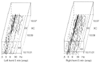 Figure 38-7
This Lifescan printout was obtained approximately 3 minutes
after occlusion (XC) of the left carotid artery. It shows attenuation of activity
on the left with occlusion. The electroencephalogram showed a similar change. The
regional cerebral blood flow with occlusion was 9 mL/100 g/min. (From Spackman
TN, Faust RJ, Cucchiara RF, et al: A comparison of a periodic analysis of the EEG
with standard EEG and cerebral blood flow for detection of ischemia. Anesthesiology
66:229, 1987.)
Figure 38-7
This Lifescan printout was obtained approximately 3 minutes
after occlusion (XC) of the left carotid artery. It shows attenuation of activity
on the left with occlusion. The electroencephalogram showed a similar change. The
regional cerebral blood flow with occlusion was 9 mL/100 g/min. (From Spackman
TN, Faust RJ, Cucchiara RF, et al: A comparison of a periodic analysis of the EEG
with standard EEG and cerebral blood flow for detection of ischemia. Anesthesiology
66:229, 1987.)
Electroencephalographic monitoring does have limitations. Despite the data available from the Mayo Clinic, immediately evident postoperative strokes have been reported in patients without any evidence of electroencephalographic changes intraoperatively. In my (M.E.M.) experience, such strokes result from emboli through the lenticulostriate arteries to subcortical structures that do not generate or affect the EEG but are critically involved in the descending motor pathway. Overall, however, such events are rare.
|
|
|
|
|
|
|
|
|
|
|
|
|