ANESTHESIA AND THE ELECTROENCEPHALOGRAM
With the advent of the then high-tech EEG in 1950, Courtin and
colleagues[22]
sought to monitor the brain and devise
a servocontrolled system that could adjust the anesthetic concentration administered
on the basis of the electroencephalographic pattern. It was an ingenious idea, but
despite available descriptions of the EEG during anesthesia, knowledge and technology
were not adequate at that time. Even using more modern devices with much higher
degrees of computer power, servocontrolled administration of anesthetics remain imperfect
at best.[23]
[24]
Because all anesthetic drugs do not produce exactly the same changes in electroencephalographic
pattern as anesthesia deepens, generic correlation of the EEG with depth of anesthesia
across all anesthetic techniques remains an elusive goal. One of the major reasons
that the EEG has been difficult to use for assessing anesthetic depth is that most
modern anesthetics use many different classes of drugs, all of which have significant
electroencephalographic effects, for premedication, induction, and
maintenance of anesthesia. Other intraoperative factors may affect the EEG ( Table
38-2
), adding to the difficulty of its interpretation.
Much research has been directed at developing a processed electroencephalographic
parameter that is indicative of depth of anesthesia. These processed parameters
fall into two broad categories: parameters derived from power analysis of the raw
electroencephalographic data and parameters derived from bispectral analysis of the
EEG. Power analysis processes data from the raw EEG related to frequency and amplitude
of the waveforms over time but does not consider phase relationships between the
component waves. Bispectral analysis is a somewhat more complex process that includes
phase relationship data. Several processed electroencephalographic parameters from
Fourier analysis have been investigated as indicators of anesthetic depth. These
parameters include spectral edge frequency 95% (SEF95),
which is the frequency below which 95% of the electroencephalographic activity falls;
median frequency (i.e., frequency at the midpoint
of the power spectrum); peak power frequency (i.e.,
frequency with the highest electroencephalographic power), and relative
delta power (i.e., percent of the EEG power in the delta band). Some
investigators have had success in using one or more of these parameters to predict
the depth of anesthesia.[14]
Others have found
that although these parameters change during anesthesia, they were not consistently
predictive of depth of anesthesia as assessed by response to stimuli, movement, or
return of consciousness during emergence from anesthesia.[25]
[26]
In particular, these parameters depend on
the
type of anesthetic agent or combination of agents used. Whereas one parameter may
correlate well with a primarily volatile anesthetic technique, it may perform less
consistently during narcotic-based anesthesia.
Encouraging results have been obtained using the BIS to monitor
anesthetic depth (see Chapter 31
).
The BIS is a processed parameter derived from multiple features generated by bispectral
analysis of the EEG. Through clinical trials, features of the bispectral analysis
that were predictive of response to stimuli under the effects of a variety of anesthetic
agents were identified. These features were combined to a multivariate index using
discriminant analysis.[18]
[27]
After the BIS was developed, it was further tested and empirically refined to improve
predictive ability.[27]
The BIS is displayed as
a numeric value from 0 to 100 and can be trended over time. Many clinical trials
have investigated the ability of the BIS to monitor anesthetic depth and predict
response to stimuli. Trials have been directed at determining BIS values predictive
of loss of consciousness, loss of recall, and prevention of movement in response
to surgical stimulation. The BIS value indicative of a certain level of consciousness
varies somewhat between different anesthetic techniques[27]
;
however, the ability of the BIS to predict loss of consciousness and lack of recall
during sedation has been consistently demonstrated by a number of investigators using
a variety of drugs and drug combinations.[14]
[27]
However, the ability of BIS to predict hemodynamic response to surgical stimulation
or movement in response to surgical stimulation has been less consistently demonstrated
and may depend more on the anesthetic technique.[28]
[29]
These results have suggested to some that
the
anesthetic state involves at least two different components. One is a reflection
of hypnosis and consists of loss of consciousness and recall; the BIS value is indicative
of this state. However, the obtundation of hemodynamic and movement responses to
noxious stimuli is less well predicted by the BIS and probably is mainly mediated
at the spinal cord level.[27]
BIS monitoring of
the level of sedation during an awake cranitomy with cortical mapping is shown in
Figure 38-5
.
Anesthetic drugs affect the frequency and amplitude of electroencephalographic
waveforms. Although each drug class and each specific drug has some explicit, dose-related
electroencephalographic effects ( Table
38-3
), some basic anesthesia-related electroencephalographic patterns may
be described. Subanesthetic doses of intravenous and inhaled anesthetics usually
produce an increase in frontal β activity and abolish the α activity normally
seen in the occipital leads in the awake, relaxed patient with the eyes closed.
As the patient goes to sleep with general anesthesia, the brain waves become larger
in amplitude and slower in frequency. In the frontal areas, small β activity
seen in the awake patient slows to the α range and increases in size. In combination
with the loss of the occipital α activity, this phenomenon produces the appearance
of a shift of the α activity from the posterior cortex to the anterior cortex.
Further increases in the dose of the inhalation or intravenous agent produce further
slowing of the EEG, and some agents have the capability to totally suppress electroencephalographic
activity (see Table 38-3
).
Other agents (e.g., opioids, benzodiazepines) never produce burst suppression or
an isoelectric EEG despite increasing dose because they are incapable of completely
suppressing the EEG or because cardiovascular toxicity of the drug (e.g., halothane)
prevents administration of a large enough dose.
Intravenous Anesthetics
Barbiturates, Propofol, and Etomidate
Despite widely varying potencies and durations of action, all
of the intravenous anesthetics produce similar electroencephalographic patterns.
Figure 38-6
shows the electroencephalographic
effects of thiopental (see Chapter
10
). These drugs all follow the basic anesthesia-related electroencephalographic
pattern described previously, with
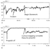 Figure 38-5
Bispectral index (BIS) tracings from an awake craniotomy
with intraoperative cortical mapping. Propofol infusion was titrated against the
BIS value to provide adequate sedation during the operative procedure. Termination
of the propofol infusion during the cortical mapping is followed by a prompt increase
in the BIS value and recovery of the patient to a fully alert state.
Figure 38-5
Bispectral index (BIS) tracings from an awake craniotomy
with intraoperative cortical mapping. Propofol infusion was titrated against the
BIS value to provide adequate sedation during the operative procedure. Termination
of the propofol infusion during the cortical mapping is followed by a prompt increase
in the BIS value and recovery of the patient to a fully alert state.
initial activation (see Fig. 38-6A
)
followed by dose-related depression. As the patient loses consciousness, characteristic
frontal spindles are seen (see Fig.
38-6B
), which are replaced by polymorphic 1- to 3-Hz activity (see Fig.
38-6C
) as the drug dose is increased. Further increases in dose result
in lengthening periods of suppression interspersed with periods of activity (i.e.,
burst suppression). With a very high dose, electroencephalographic silence results.
All these drugs have been reported to cause epileptiform activity in humans, but
epileptiform activity is clinically significant only after methohexital and etomidate
when given in subhypnotic doses.
Ketamine
Ketamine does not follow the basic anesthesia-related electroencephalographic
pattern. Anesthesia with ketamine is characterized by frontally dominant rhythmic,
high-amplitude theta activity. Increasing doses produce intermittent polymorphic
δ activity of very large amplitude interspersed with low-amplitude β activity.
[30]
Electrocortical silence cannot be produced
with ketamine. Electroencephalographic activity may be very disorganized and variable
at all doses. Recovery of normal electroencephalographic activity, even after a
single bolus dose of ketamine, is relatively slow compared with barbiturates. There
is no information available about the relationship between emergence reactions after
ketamine and the EEG. Ketamine has also been associated with increased epileptiform
activity.[30]
Benzodiazepines
Despite different potencies and durations of actions, benzodiazepines
also follow the basic anesthesia-related electroencephalographic pattern. As a class,
however, these drugs are incapable of producing burst suppression or an isoelectric
EEG.
Opioids
As a class, opioids do not follow the basic anesthesia-related
electroencephalographic pattern. In general, opioids produce a dose-related decrease
in frequency and increase in amplitude of the EEG. If no further doses of opiates
are given, α and β activity will return as drug redistribution occurs.
The rapidity of return depends on the initial dose and on the drug. Remifentanil
is associated with the most rapid return to normal.[31]
Complete suppression of the EEG cannot be obtained with the opioids. Epileptiform
activity occurs in humans and in animals receiving large to supraximal clinical doses
of opioids. For example, sharp wave activity is relatively common after induction
of anesthesia with fentanyl, with 20% of patients showing this phenomenon after 30
µg/kg, 60% at 50 µg/kg, 58% at 60 µg/kg, and 80% at 70 µg/kg.
This epileptiform activity is mainly observed in the frontotemporal region.[32]