MONITORING OF HIGH-FREQUENCY VENTILATION
High-frequency ventilation has been variously defined but in general
represents mechanical ventilation at high rates (usually 160 breaths/min). Several
means of ventilating in this manner have been used experimentally and clinically.
The use of conventional positive-pressure ventilators at high rates and small tidal
volumes is called high-frequency positive-pressure ventilation
(HFPV). Tidal volumes are usually on the order of 3 to 4 mL/kg of body weight, with
a frequency of 60 to 100 breaths/min. The use of an oscillator providing positive
and negative pressure fluctuations (e.g., a loudspeaker) is called high-frequency
oscillatory ventilation (HFOV). Higher frequencies, upward of 3000 cycles/min,
have been used with this modality. A bias flow of
fresh gas at the level of the oscillator provides the source of the respiratory gas
and washes out CO2
. Injection of a high-velocity pulse of gas into the
airway through a narrow cannula, entraining with it fresh gas, is called high-frequency
jet ventilation (HFJV).
In all these forms of high-frequency ventilation, instantaneous
gas flows and pressure fluctuations cannot
usually be monitored with conventional transducers. Moreover, because the system
is basically open, a portion of the gas flow directed into the airway may leak out
and not participate in intrapulmonary gas exchange. High-frequency ventilatory fluctuations
generated by these ventilators may also in part do nothing more than compress and
decompress the compliance of the ventilatory circuit and large conducting airways.
Conventional mechanical monitoring is therefore difficult. Capnography is difficult
to apply, because dilution of expired gas may render endtidal measurements artificially
low, even assuming a high-fidelity, high-frequency capnograph.
Monitoring of patients receiving high-frequency ventilation requires
the ability to monitor O2
and CO2
exchange, as well as mechanical
safety, including airway disconnection and obstruction. Hoskyns and colleagues[257]
measured tidal volumes in 0.8- to 1.9-kg infants ventilated at 2 to 25 Hz using an
external respiratory jacket. A side port of the jacket was used to monitor pressure
changes, which correlated linearly with tidal volume.
Whereas patient oxygenation can readily be monitored with pulse
oximetry, there is not reliable noninvasive monitor of CO2
exchange.
One way of monitoring CO2
is to measure the mean waste gas CO2
concentration by placing a capnograph in the expired circuit. If any condition that
interferes with CO2
exchange develops, the mean expired CO2
decreases. although this method provides a fairly gross measure of adequacy of CO2
exchange, the expired CO2
concentration highly depends on fresh gas flow
rate. A more satisfactory monitor would multiply fresh gas flow by expired CO2
fraction to obtain V̇CO2
. Changes
in CO2
could then reflect a mechanical problem with the ventilator. Unfortunately,
other factors such as anesthesia or hypothermia may alter V̇CO2
.
The clinician cannot obtain a measure of PACO2
or PaCO2
by this method. The most commonly
used method is to interject a conventional breath periodically to measure end-tidal
CO2
.[258]
Capnometry can be used to
measure end-tidal CO2
during HFJV without such a maneuver, provided that
gas is sampled from a port situated at the tip of the endotracheal tube.[259]
Monitoring of airway pressure is extremely important in high-frequency
ventilation. In particular, HFJV uses high pressures and gas flows. Expiratory
port occlusion can therefore result in extremely high airway pressures. Gas pressures
commonly are measured on both sides of the jet valve (i.e., drive pressure and jet
pressure), along with an independent pressure measurement in the airway ( Fig.
36-25
). An automated feedback loop is required to interrupt the jet ventilation
by closing the solenoid valve in the event of excessively high pressures in the airway
or on the jet side of the valve. Low pressures can be used as indications of airway
disconnection or ventilator malfunction.
In addition to safety concerns, airway pressure has been shown
in several studies to correlate with gas exchange efficiency during HFJV.[260]
Increasing peak airway pressures result in lower PaCO2
.
A superior indicator of PaCO2
is the
difference between peak airway pressure and end-expiratory airway pressure.[261]
However, there is no unique relationship, and the PaCO2
obtained for a given patient depends on properties of the lung. Position of the
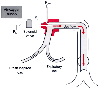 Figure 36-25
High-frequency jet ventilation. The jet is created when
a high-pressure air-O2
supply is rapidly modulated by the solenoid valve.
Fresh inspired gas is from a low-pressure source, typically an anesthesia circuit.
Drive pressure (PD) and jet pressure (PJ)
are customarily monitored to detect solenoid or jet malfunction. An independent
monitor of airway pressure (Paw), which can reliably detect overpressurization of
the airway, circuit disconnection, or ventilator malfunction, should also be available.
Figure 36-25
High-frequency jet ventilation. The jet is created when
a high-pressure air-O2
supply is rapidly modulated by the solenoid valve.
Fresh inspired gas is from a low-pressure source, typically an anesthesia circuit.
Drive pressure (PD) and jet pressure (PJ)
are customarily monitored to detect solenoid or jet malfunction. An independent
monitor of airway pressure (Paw), which can reliably detect overpressurization of
the airway, circuit disconnection, or ventilator malfunction, should also be available.
monitoring transducer may be critical because proximal airway pressures may be artifactually
low.[261]
Jet ventilation for prolonged periods should ideally be performed
on patients with the ability to monitor arterial blood gases directly. Periodic
measurement of PaCO2
may provide greater
assurance of adequate pulmonary gas exchange than simple reliance on noninvasive
measures.