|
|
|
|
|
|
|
|
|
|
|
|
|
|
|
Estimates of compliance and resistance in the respiratory system may be obtained by inspection of the airway pressure-volume (or time) relationships. Thoracic compliance may be obtained by inflating the lungs with known increments of volume and measuring the static pressure at each point. Under normal circumstances, the lung and chest wall have approximately equal compliance of approximately 200 mL/cm H2 O, resulting in a total thoracic compliance of 100 mL/cm H2 O. Individual measurements of chest wall and lung components of the total respiratory compliance cannot be made without some estimate of pleural pressure. Conventionally, pleural pressure is measured by inserting a pressure-monitoring balloon within the esophagus. Unfortunately, in the supine patient, the weight of the mediastinum results in a falsely high estimate of pleural pressure when using this technique. Direct pleural pressure measurement must therefore be obtained to measure lung and chest wall compliance accurately in this position. A noninvasive method of pleural pressure measurement, using a flat loop of Teflon-insulated wire attached to the skin of the suprasternal fossa, has been described.[228] The skin movement that accompanies changes in intrapleural pressure is transduced by the coil of wire by continuous measurement of its self-inductance. Measurements of pulmonary compliance using this technique correlated well with similar measurements using an esophageal balloon to estimate intrapleural pressure. However, the technique is time consuming, and measurements cannot be obtained in some individuals.
Thoracic compliance can be estimated on a breath-by-breath basis by monitoring the inspiratory pressure-time curve. If a constant tidal volume is being used to ventilate a patient, the thoracic compliance will be inversely related to the inspiratory pressure. It is important that the pressure used to calculate thoracic compliance be at end inspiration (i.e., plateau pressure), because during the period of inspiratory gas flow there is an additional component of pressure due to airways resistance. The changes in peak airway pressure (related to airways resistance and
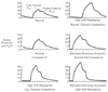 Figure 36-22
Examples of airway pressure waveforms. Airway pressure
is plotted versus time. Decreased static thoracic compliance results in an increase
in plateau pressure; increases in airway resistance cause increased peak airway pressure
and decreased dynamic compliance.
Figure 36-22
Examples of airway pressure waveforms. Airway pressure
is plotted versus time. Decreased static thoracic compliance results in an increase
in plateau pressure; increases in airway resistance cause increased peak airway pressure
and decreased dynamic compliance.
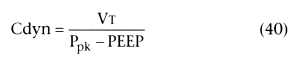
Static compliance (Cstat) can be calculated by measuring the plateau
pressure and using the following equation:
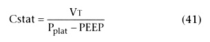
The normal value for static compliance is 50 to 100 mL/cm H2
O.
To make these compliance calculations, any end-expiratory pressure must be subtracted from the peak pressure or plateau pressure. The PEEP value can usually be read directly from the airway pressure monitor at end expiration. However, when insufficient expiratory time prevents complete emptying of the lungs, significant end-expiratory elevation of alveolar pressure that is not detected at the airway pressure monitor (i.e., auto-PEEP) may occur.[229] [230] Auto-PEEP is most likely to exist when elevation of airway resistance or compliance cause prolongation of expiratory time or when high ventilation rates are required.[229] Auto-PEEP can result in decreased cardiac output, hypotension, and electromechanical dissociation in mechanically ventilated patients. It also affects compliance measurements. Detection of auto-PEEP can be accomplished by occluding the exhalation port of the ventilator while delaying the onset of the next ventilator breath. Alveolar pressure then equilibrates with the ventilator circuit, and auto-PEEP can be read on the airway pressure monitor ( Fig. 36-23 ). During this temporary occlusion maneuver, it is important to prevent supplementary gas flow (e.g., fresh gas flow) from entering the circuit and falsely elevating the auto-PEEP value.
A decrease in static compliance may be caused by atelectasis, pulmonary edema, pneumothorax, external compression of the chest (i.e., intra-abdominal hemorrhage or, in the operating room, abdominal packing or the surgeon's leaning on the torso), or accumulation of pleural fluid. Decreased dynamic compliance may be caused by elevated airways resistance (e.g., bronchospasm, mucus accumulation) or obstruction or kinking of the endotracheal tube.
In parallel with the patient's thoracic compliance are the internal compliances of the breathing circuit and any associated humidification or gas warming systems. Circuit compliance may be as high as 10 mL/cm H2 O and must be accounted for if an accurate measurement of thoracic compliance is required. Decreased thoracic compliance places an additional load on the patient's respiratory muscles. Thoracic compliance less than 25 mL/cm H2 O is unlikely to result in successful weaning from mechanical ventilation.[231]
Thoracic resistance can be measured in the intubated patient by oscillating the airway with a sinusoidal or randomly varying flow with simultaneous measurements of flow and pressure. Significant increases in thoracic resistance have been observed in anesthetized patients after reversal of neuromuscular blockade with edrophonium (0.43 mg/kg).[232] An indirect estimate of airways resistance may also be obtained by monitoring the flow-time
 Figure 36-23
Auto-positive end-expiratory pressure (auto-PEEP). Severe
airway obstruction may result in high alveolar gas pressure at end expiration, which
can exert the same effect on hemodynamics as externally applied PEEP. This is not
usually detectable unless the expiratory port of the ventilator circuit is occluded.
Auto-PEEP may then be read directly from the airway pressure monitor. (Adapted
from Pepe PE, Marini JJ: Occult positive end-expiratory pressure in mechanically
ventilated patients with airflow obstruction: The auto-PEEP effect. Am Rev Respir
Dis 126:166, 1982.)
Figure 36-23
Auto-positive end-expiratory pressure (auto-PEEP). Severe
airway obstruction may result in high alveolar gas pressure at end expiration, which
can exert the same effect on hemodynamics as externally applied PEEP. This is not
usually detectable unless the expiratory port of the ventilator circuit is occluded.
Auto-PEEP may then be read directly from the airway pressure monitor. (Adapted
from Pepe PE, Marini JJ: Occult positive end-expiratory pressure in mechanically
ventilated patients with airflow obstruction: The auto-PEEP effect. Am Rev Respir
Dis 126:166, 1982.)
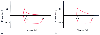 Figure 36-24
Flow (ordinate) versus volume (abscissa). A,
Closed-chest positive-pressure ventilation under general anesthesia in a patient
with severe airways obstruction and hyperinflation before surgery to reduce lung
volume. The flow-volume curve shows inspiratory (negative) and expiratory (positive)
flow on the ordinate, plotted clockwise from zero volume on the abscissa. Expiratory
flow started with a sharp upward peak and then fell immediately to a low flow rate
with convexity toward the volume axis, suggesting expiratory flow limitation. expiratory
flow rate was so low that inflation of the next positive-pressure breath was initiated
before expiratory flow reached zero. Because expiratory flow continued up to this
point, there must have been intrinsic positive end-expiratory pressure (PEEPi).
B, A similar closed-check flow-volume curve after
lung resection shows that the characteristic pattern of expiratory flow limitation
has disappeared and that expiratory flow rate fell to zero before inflation started
for the next breath (i.e., no suggestion of PEEPi). (Adapted from Dueck
R: Assessment and monitoring of flow limitation and other parameters from flow/volume
loops. J Clin Monit Comput 16:425, 2000.)
Figure 36-24
Flow (ordinate) versus volume (abscissa). A,
Closed-chest positive-pressure ventilation under general anesthesia in a patient
with severe airways obstruction and hyperinflation before surgery to reduce lung
volume. The flow-volume curve shows inspiratory (negative) and expiratory (positive)
flow on the ordinate, plotted clockwise from zero volume on the abscissa. Expiratory
flow started with a sharp upward peak and then fell immediately to a low flow rate
with convexity toward the volume axis, suggesting expiratory flow limitation. expiratory
flow rate was so low that inflation of the next positive-pressure breath was initiated
before expiratory flow reached zero. Because expiratory flow continued up to this
point, there must have been intrinsic positive end-expiratory pressure (PEEPi).
B, A similar closed-check flow-volume curve after
lung resection shows that the characteristic pattern of expiratory flow limitation
has disappeared and that expiratory flow rate fell to zero before inflation started
for the next breath (i.e., no suggestion of PEEPi). (Adapted from Dueck
R: Assessment and monitoring of flow limitation and other parameters from flow/volume
loops. J Clin Monit Comput 16:425, 2000.)
Commercially available monitors can provide continuous displays of inspired volume versus airways pressure and flow versus inspired volume[234] [235] ( Fig. 36-24 ). These techniques can been used to monitor changes in pulmonary compliance caused by intraoperative pulmonary edema[234] or during laparoscopic instillation of gas into the peritoneal cavity,[236] to detect malpositioning of doublelumen endotracheal tubes,[237] and to assess pulmonary mechanics after lung volume-reduction surgery.[235]
|
|
|
|
|
|
|
|
|
|
|
|
|