|
|
|
|
|
|
|
|
|
|
|
|
|
|
|
One of the major objections to the use of ear oximeters is the required assumption that the earlobe contains predominantly arterial blood, because these instruments had no means of differentiating between arterial and venous hemoglobin. The pulse oximeter is able to make this differentiation by assuming that the pulsatile portion of the signal is entirely arterial blood. This is almost always true except under unusual clinical circumstances, such as when there are prominent venous pulsations as in tricuspid regurgitation.[59]
The principle of pulse oximetry is shown schematically in Figure
36-10
. The light passing through tissue is absorbed by tissue and by venous
and arterial blood. The ratio S is calculated at two wavelengths of light, usually
around 660 nm (red) and 940 nm (infrared), for which the following relationships
apply:
In Equation 21, AC660
and AC940
are pulsatile components of
absorbance at wavelengths of 660 and 940 nm, respectively. DC660
and
DC940
are the corresponding steady-state components. S is empirically
related to O2
saturation and incorporated into the design of the instrument.
It has been proposed that the assessment of SaO2
by pulse oximetry be designated as SpO2
.
[50]
In practice, pulse oximeters use two light-emitting diodes (LEDs) and one photo diode as transmitting and sensing transducers, usually placed on opposite sides of a digit. The two LEDs are activated alternatively. The ratio S is calculated electronically; from the value of S, SpO2 is derived by an internally stored algorithm.
One drawback of oximetry is that it is rather insensitive to large changes in arterial PO2 at the high end of the Hb-O2 dissociation curve, where large changes in PO2 are associated with small changes in SpO2 . Another problem is that because only two wavelengths are used, only two species of hemoglobin can be resolved (assumed to be
 Figure 36-10
Principle of pulse oximetry. Light passing through tissue
containing blood is absorbed by tissue and by arterial, capillary, and venous blood.
Usually, only the arterial blood is pulsatile. Light absorption may therefore be
split into a pulsatile component (AC) and a constant or nonpulsatile component (DC).
Hemoglobin O2
saturation may be obtained by application of Equation 19
in the text. (Data from Tremper KK, Barker SJ: Pulse oximetry. Anesthesiology
70:98, 1989.)
Figure 36-10
Principle of pulse oximetry. Light passing through tissue
containing blood is absorbed by tissue and by arterial, capillary, and venous blood.
Usually, only the arterial blood is pulsatile. Light absorption may therefore be
split into a pulsatile component (AC) and a constant or nonpulsatile component (DC).
Hemoglobin O2
saturation may be obtained by application of Equation 19
in the text. (Data from Tremper KK, Barker SJ: Pulse oximetry. Anesthesiology
70:98, 1989.)
Although the inherently reliability of pulse oximetry has led to its wide use in anesthesia and critical care, remaining problems include motion sensitivity, causing false alarms and erroneous measurements, and hypoperfusion, causing loss of signal (see Chapter 30 ). Several manufacturers have developed proprietary methods to address these problems based on analysis of frequency, waveform morphology, or saturation. [66] [67]
Published evidence supports the ability of new generation pulse oximeters to detect hypoxemic episodes more reliably than conventional devices under conditions of patient motion[68] and hypothermic hypoperfusion,[69] although they often falsely indicate hypoxemia, particularly when intra-aortic balloon pumps are used.[70]
To assess arterial oxygenation when it is impossible to provide a transmission path, reflectance oximetry has been exploited. Reflectance oximetry has been used to monitor fetal scalp during labor.[71] An esophageal probe has been developed to monitor SpO2 when extremities are unavailable or have a pulse of insufficient amplitude.[72] Monitors using forehead skin probes are also commercially available.[73] [74] Forehead sensors appear less susceptible to motion artifact during exercise, and during
 Figure 36-11
Effect of pulse oximeter probe replacement on delay from
onset of hypoxemia to a drop in the measured SpO2
.
During cold-induced peripheral vasoconstriction in normal volunteers, the onset
of hypoxemia was detected more quickly using an oximeter probe on the forehead compared
with the finger. Other studies have shown a similar advantage for pulse oximeter
probes placed on the ear. (From Bebout DE, Mannheimer PD, Wun C-C: Site-dependent
differences in the time to detect changes in saturation during low perfusion. Crit
Care Med 29:A115, 2002.)
Figure 36-11
Effect of pulse oximeter probe replacement on delay from
onset of hypoxemia to a drop in the measured SpO2
.
During cold-induced peripheral vasoconstriction in normal volunteers, the onset
of hypoxemia was detected more quickly using an oximeter probe on the forehead compared
with the finger. Other studies have shown a similar advantage for pulse oximeter
probes placed on the ear. (From Bebout DE, Mannheimer PD, Wun C-C: Site-dependent
differences in the time to detect changes in saturation during low perfusion. Crit
Care Med 29:A115, 2002.)
Current pulse oximeters use only two wavelengths and can resolve only two hemoglobin species. Use of additional wavelengths provides the possibility of measuring additional hemoglobins. There are no commercially available devices, although research in this area is ongoing.
Although oximetry has been available since the 1940s, the development of pulse oximetry triggered its wide acceptance because of the latter's reliability and convenience. The reliability and ease of use of such monitors has led to incorporation of their use within standards of care. Pulse oximetry has become a standard component of anesthesia monitoring, and it has gained acceptance in postanesthesia care units (PACUs) and intensive care units (ICUs) and for patients undergoing a variety of diagnostic procedures such as gastrointestinal endoscopy. The wide application of pulse oximetry attests to the clinical utility of this major advance in monitoring.
In a trial in which 20,802 patients scheduled for surgery were randomly assigned to receive monitoring with pulse oximetry or not, hypoxemia was detected during anesthesia 20 times and hypoventilation three times as frequently in the pulse oximetry group.[76] [77] These differences persisted in the PACU, and bronchospasm, atelectasis, and bradycardia also were detected more frequently in the group monitored with oximetry. On the basis of favorable clinical experience with this technology, pulse oximetry is now a standard monitoring technique during anesthesia and monitored sedation.
In patients admitted to an ICU after elective cardiac surgery, the use of pulse oximetry has been reported to reduce the number of arterial blood gas analyses and to increase the probability of detecting hypoxemic episodes.[78] Continuous monitoring of patients for up to several days after surgery has been reported to detect hypoxemic episodes.[79] [80] [81] [82] [83] [84] [85] [86] [87] [88] [89] [90] [91] [92] [93] [94] In this setting, some of the low readings can be caused by motion artifact, the elimination of which requires appropriate methods.[95]
These studies indicate that significant desaturation (SpO2 of 80% to 85%) is relatively common. Desaturation also occurs in some patients before surgery,[86] [87] [89] [90] and there is evidence that the existence of preoperative hypoxemic episodes may predict desaturation postoperatively. Hypoxemia is often correlated with peak effects of analgesia.[84] [91] Detection or prevention of such episodes may be beneficial, because arterial desaturation can be correlated with an increase in heart rate[83] [85] and electrocardio-graphic evidence of myocardial ischemia.[85] [88]
Because pulse oximeters are dual-wavelength devices, the presence of hemoglobin species other than HHb and HbO2 must therefore result in erroneous readings ( Table 36-4 ).
The effect of HbCO may be discerned by examining its absorption
spectrum. At 920 nm, HbCO has an extremely low absorbance and therefore does not
contribute to total absorbance. At 660 nm, however, HbCO has an absorbance very
similar to that of HbO2
, and SpO2
will therefore be falsely high. This effect has been measured in a canine study
in which the pulse oximeter reading of Nellcor and Ohmeda pulse oximeters was approximated
[96]
by the following formula:
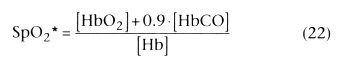
In Equation 22, SpO2
* is the pulse
oximeter reading.
Using a pulse oximeter in the presence of HbCO provides a falsely high estimate of arterial SO2 . In that study, when HbCO was 50%, SpO2 was approximately 95%. In human cases of carbon monoxide poisoning, a similar error has been observed. [97] [98] [99] [100] [101] In 30 patients with HbCO levels ranging from 25.2% to 54% (mean, 36.9%), Hampson[101] observed that each 8% increase in HbCO resulted in only a 1% decrease in SpO2 reading.
HbMet has a larger absorbance than either of the two major species of hemoglobin at 940 nm but simulates hemoglobin at 660 nm. At high SaO2 levels (>85%) the reading underestimates the true value; at a low SaO2 (<85%), the value is falsely high.[102] [103] In the presence of high HbMet concentrations, the measured SpO2 approaches 85%, independently of the actual arterial oxygenation.
Sulfhemoglobin, which can form after exposure to certain drugs and chemicals, and cyanmethemoglobin, a product of the pharmacologic induction of methemoglobinemia for treatment of cyanide poisoning, are nonfunctional hemoglobins. Although sulfhemoglobinemia is known to produce errors in CO oximetry,[104] [105] [106] usually with a false reading of methemoglobin, the effects of these species on pulse oximetry have not been reported.
In neonates, there is a large proportion of HbF. Because HbF has almost the same absorption spectrum as hemoglobin A, it has almost no measurable effect on SpO2 .[63]
SpO2 has been reported to be accurate in patients with hemoglobin S (HbS) disease when SaO2 determined by this method was compared with SaO2 obtained by using in vitro oximetry,[107] [108] [109] although it is possible that the presence of HbS produces a similar artifact in both types of instrument. During sickle cell crisis, it has been reported that SpO2 overestimates SaO2 measured by CO oximetry [110] by an average of 6.9%. Patients with sickle cell disease have a rightward shift of the Hb-O2 dissociation curve (i.e., increased P50 ), and at any given PaO2 value, the SpO2 is lower than the normal Hb-O2 dissociation curve would predict.
Pulse oximetry evaluation has been reported in a patient with hemoglobin H anemia. Two different pulse oximeters read 93% and 95% (artifactually high), respectively, in a patient who had a hematocrit of 9%.[111] Independent measurements of SaO2 with which to compare the pulse oximeter readings were not reported.
Hemoglobin Köln, an unstable hemoglobin, has been associated with an artifactual reduction of 8% to 10% in SpO2 reading.[112] [113]
Because of changes in their optical absorption spectra, hemoglobin substitutes could affect the readings of pulse oximeters or CO oximeters (see Chapter 47 ). Appropriate testing would have to incorporate independent measurement of O2 content; however, little information exists on the effects of hemoglobin substitutes on CO oximetry or pulse oximetry. Artificial hemoglobin solutions such as diaspirin-cross-linked hemoglobin[114] and bovine polymerized hemoglobin (oxygen carrier-201)[115] [116] have no apparent effect on SpO2 , at least after infusion of relatively low doses and under normoxic conditions. Whether administration of these solutions may artifactually increase SpO2 readings has not been firmly established. Some errors are likely. An in vitro study of five hemoglobin substitutes suggested that CO oximeters could provide clinically acceptable measurements of total hemoglobin concentration, although measurement of components (e.g., methemoglobin) might be inaccurate.[117] In dogs, hemoglobin glutamer-200 administration (Oxyglobin, Biopure, Cambridge, MA) does not appear to affect significantly the readings of one commercially available CO oximeter (Nova Biomedical, Waltham MA).[118] The availability of a 128-nm-wavelength CO oximeter offers the potential for resolving HbF, sulfhemoglobin, and artificial blood substitutes.[119]
At normal oxygenation levels in humans, over a range of hemoglobin levels from 2.3 to 8.7 g/dL, SpO2 accurately
| Factor | Effect | References |
|---|---|---|
| Toxic Alterations in Hemoglobin | ||
| Carboxyhemoglobin (COHb) | Slight reduction of the assessment of SaO2 by pulse oximetry (SpO2 ) (i.e., overestimates fraction of Hb available for O2 transport) | [97] [98] [99] [100] [101] |
| Cyanmethemoglobin | Not reported |
|
| Methemoglobin (MetHb) | At high levels of MetHb, SpO2 approaches 85%, independent of actual oxygen saturation (SaO2 ) | [102] [103] |
| Sulfhemoglobin | Not reported (affects CO oximetry by producing a falsely high reading of MetHb) | [104] [105] [106] |
| Structural Hemoglobinopathies | ||
| Hemoglobin F | No significant effect | [63] |
| Hemoglobin H | No significant effect (i.e., overestimates fraction of Hb available for O2 transport) | [111] |
| Hemoglobin Köln | Artifactual reduction in SpO2 of 8–10% | [112] [113] |
| Hemoglobin S | No significant effect |
|
| Hemoglobin Replacement Solutions | ||
| Diaspirin cross-linked hemoglobin | No significant effect | [114] |
| Bovine polymerized hemoglobin (oxygen carrier-201) | No significant effect | [115] [116] |
| Dyes | ||
| Fluorescein | No significant effect | [62] [125] |
| Indigo carmine | Transient decrease |
|
| Indocyanine green | Transient decrease | [62] [125] |
| Isosulfan blue (patent blue V) | No significant effect at low dose; prolonged reduction in SpO2 at high dose | [130] [131] [132] |
| Methylene blue | Transient, marked decrease in SpO2 , lasting up to several minutes; possible secondary effects due to effects on hemodynamics | [124] [125] [126] [127] [128] |
| Hemoglobin Concentration | ||
| Anemia | If SaO2 normal: no effect; during hypoxemia, at Hb values less than 14.5 g/dL: progressive underestimation of actual SaO2 | [120] [121] |
| Polycythemia | No significant effect | [122] [123] |
| Other Factors | ||
| Acrylic fingernails | No significant effect |
|
| Ambient light interference | Bright light, particularly if flicker frequency is close to a harmonic of light-emitting diode switching frequency, can falsely elevate SpO2 reading. | [64] [142] |
| Arterial O2 saturation | Depends on manufacturer. During hypoxemia, SpO2 tends to be artifactually low. | [5] [73] [141] [303] |
| Blood flow | Reduced amplitude of pulsations can hinder obtaining a reading or cause a falsely low reading. | [146] [147] |
| Henna | Red henna: no effect; black henna: may block light sufficiently to preclude measurement | [134] |
| Jaundice | No effect. Multi-wavelength laboratory oximeters may register a falsely low SaO2 and a falsely high COHb and MetHb. | [135] [136] [137] [138] |
| Motion | Movement, especially shivering, may depress SpO2 reading. | [64] |
| Nail polish | Slight decrease in SpO2 reading, with greatest effect using blue nail polish, or no change | [133] [304] |
| Sensor contact | "Optical shunting" of light from source to detector directly or by reflection from skin results in falsely low SpO2 reading. | [145] |
| Skin pigmentation | Small errors or no significant effect reported. Deep pigmentation can result in reduced signal. | [62] |
| Tape | Transparent tape between sensor and skin has little effect. Falsely low SpO2 has been reported when smeared adhesive is in the optical path. | [139] [140] |
| Vasodilatation | Slight decrease | [148] |
| Venous pulsation (e.g., tricuspid insufficiency) | Artifactual decrease in SpO2 | [59] |
Polycythemia has no apparent effect on pulse oximeter reading. In children with cyanotic congenital heart disease, many of whom were polycythemic, there was no systematic error in the SaO2 reading that could be attributed to high hemoglobin concentration.[122] [123]
Clinically used dyes may also have an effect on pulse oximetry.
Methylene blue results in a severe decrease in measured SaO2
.
[124]
[125]
[126]
[127]
[128]
Its
administration can also cause alterations in cardiac output (i.e., an increase and
then a decrease). In the presence of V̇A/![]() mismatch, this may result in a transient increase and then a decrease in actual SaO2
.
The presence of high concentrations of methylene blue in blood can also alter CO
oximeter readings such that an artifactual decrease in measured O2
saturation
may result. Quantitative information, however, is lacking.
mismatch, this may result in a transient increase and then a decrease in actual SaO2
.
The presence of high concentrations of methylene blue in blood can also alter CO
oximeter readings such that an artifactual decrease in measured O2
saturation
may result. Quantitative information, however, is lacking.
Indocyanine green causes less artifactual decrease than methylene blue,[125] and a still smaller decrease occurs with indigo carmine.[62] [125] Fluorescein injection has no measurable effect.[62] [125]
Isosulfan blue (i.e., sulfan blue or patent blue V), a synthetic coal tar dye used to visualize lymphatic vessels for surgical procedures, has a peak absorption[129] at 640 nm and increased absorbance at 660 nm. Its administration has been associated with prolonged artifactual reduction in SpO2 .[130] [131] [132] It tends to affect CO oximetry by producing an artifactual increase in methemoglobin and a negative carboxyhemoglobin reading. This phenomenon is not observed with subcutaneous injection of up to 100 mg into an adult.
Effects of nail polish have also been measured. Blue nail polish, with absorbance near 660 nm, has the greatest effect, an artifactual decrease, on the SaO2 reading. Other colors have smaller effects.[133] Red henna, a pigment used for hand and nail decoration in some tropical countries, has no significant effect on SpO2 , although black henna can block enough light to prevent a satisfactory reading.[134]
High levels of bilirubin have no significant effect on pulse oximeter readings,[135] [136] although older types of ear oximeters may measure a falsely low value.[58] In the presence of jaundice, multiple-wavelength laboratory oximeters may also register falsely low SaO2 and falsely high HbCO and HbMet values.[137] [138]
Deeply pigmented skin can result in inability to pick up arterial pulsations by a pulse oximeter. Small errors in SaO2 readings from black individuals have been reported by some investigators, with others finding no significant effect.[62]
Four varieties of transparent tape have been shown not to alter SpO2 measurements between 85% and 100%. [139] However, smeared adhesive caused by reusing a disposable oximeter probe has been reported to cause falsely low SpO2 readings.[140]
Practical difficulties are associated with in vivo calibration of pulse oximeters in humans at low SaO2 values. Using normal volunteers and direct measurement of SaO2 in arterial blood, Severinghaus and associates[141] compared SpO2 obtained from a variety of pulse oximeters during brief periods of profound hypoxemia. The mean nadir of arterial hemoglobin O2 saturation was 56%. Within the pulse oximeters tested at that time, there was considerable variation. At 75% SaO2 , the bias (i.e., systematic error) was scattered uniformly around zero, with individual units either overestimating or underestimating the true SaO2 by as much as 7%. Below 60% SaO2 , most units underestimated the actual SaO2 (i.e., false low measurement).
Ambient light, particularly fluorescent light, can falsely increase the SpO2 reading,[64] [142] This occurs especially if the flicker frequency of the light is close to a harmonic of the diode switching frequency. Ambient light does not seem to affect readings when oxygenation is normal.[143] An artifact due to an optical interaction between a pulse oximeter probe and the plasma touch screen of an automated data recording system has been reported to cause an artifactual reading of 100%, even with the probe disconnected from the patient. [144] Poor contact of the sensor with the skin can result in direct "optical shunting" of light from source to detector, directly or by reflection from the skin, resulting in a falsely low SpO2 reading.[145] Reduced blood flow to the extremity results in a diminished signal and can cause inability to obtain an SpO2 reading[146] or a falsely low reading.,[146] [147] possibly caused in part by greater fractional tissue consumption of arterial O2 , resulting in a lower saturation in the pulsatile blood component that is measured. There may be a slight reduction (approximately 1%) in measured SpO2 during the reactive hyperemia that occurs after ischemia in the arms,[148] perhaps due to venous pulsations. Pulse oximetry while using an intra-aortic balloon pump can produce false indication of hypoxemia.[70]
Vasoconstriction or hypotension can result in loss of SpO2 signal. Successful countermeasures have included topical application of nitroglycerin ointment and digital nerve block is often successful. Topical application of EMLA cream (2.5% lidocaine and 2.5% prilocaine) (Astra Pharmaceuticals, Westborough, MA) to the earlobe, covered by an occlusive plastic dressing for 30 minutes, has been reported to facilitate a reliable signal.[149]
In a cold environment, studies in normal volunteers have shown delayed detection of hypoxemia and re-oxygenation when the probe is on a finger or ear compared with a reflectance forehead sensor[150] [151] (see Fig. 36-11 ).
|
|
|
|
|
|
|
|
|
|
|
|
|