MEASUREMENT OF OXYGEN SATURATION
Several species of hemoglobin may exist in blood, such as oxyhemoglobin
(HbO2
), deoxygenated hemoglobin (HHb), carboxyhemoglobin (HbCO), and methemoglobin
(HbMet). To avoid ambiguity, the following definitions have been proposed.[50]
SO2
= HbO2
/(HbO2
+ HHb) (19)
HbO2
fraction = HbO2
/Total Hb (20)
SO2
is most commonly reported as a percentage,
obtained by multiplying the value obtained from Equation 19 by 100.
Measurement of SaO2
is an alternative method to PO2
for assessing
arterial blood oxygenation. The fact that absorption and reflectance spectra of
hemoglobin are affected by its oxygenation allows for convenient optical methods
of measurement. The traditional method for monitoring SaO2
is to observe the skin and mucous membranes for cyanosis. In 1947, a systematic
comparison of the clinical detection of a cyanosis by medical staff and blood O2
saturation measurement with an ear oximeter in normal volunteers demonstrated the
poor accuracy of cyanosis as an indicator of hypoxemia.[51]
In this study, a total of 7204 observations of skin and mucous membrane color were
made in normal volunteers breathing air or hypoxic gas over a measured SaO2
range of 71% to 100%. False-positive diagnosis of cyanosis was common; cyanosis
was diagnosed in 37% of 4587 observations despite a measured SaO2
of 91% to 100%. However, in 1723 observations of hypoxic volunteers with a measured
SaO2
between 71% and 80%, normal color
was observed in 12% of cases.
Cyanosis seems to be correlated best with the quantity of deoxygenated
arterial blood. It has been suggested that for cyanosis to be detectable, 5 g/dL
of deoxygenated hemoglobin must be present in the arterial blood,[52]
although published evidence is consistent with a lower detection threshold[53]
( Fig. 36-9
). Nevertheless,
the threshold for visual detection of cyanosis depends on total hemoglobin concentration.
If 3 g/dL of desaturated blood is required to detect cyanosis by visual observation,
at a total hemoglobin concentration of 15 g/dL, cyanosis occurs when SaO2
is below 80%, compared with 66% for a hemoglobin level of 9 g/dL. The evident inaccuracy
and uncertainty inherent in attempts to assess oxygenation by visual inspection have
led to its replacement with highly successful quantitative methods.
Carbon Monoxide Oximetry
Simultaneous measurement of several hemoglobin species with different
absorption spectra can be accomplished by using multiple-wavelength absorption with
at
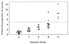 Figure 36-9
Quantity of deoxygenated hemoglobin (Hb) in arterial
blood as a function of the degree of cyanosis. Traditionally, it has been assumed
that 5 g/dL of deoxygenated hemoglobin is required for the detection of cyanosis,
but mild cyanosis can be observed at lower values. (Data from Stadie WC:
The oxygen of the arterial and venous blood in pneumonia and its relation to sepsis.
J Exp Med 30:215, 1919.)
Figure 36-9
Quantity of deoxygenated hemoglobin (Hb) in arterial
blood as a function of the degree of cyanosis. Traditionally, it has been assumed
that 5 g/dL of deoxygenated hemoglobin is required for the detection of cyanosis,
but mild cyanosis can be observed at lower values. (Data from Stadie WC:
The oxygen of the arterial and venous blood in pneumonia and its relation to sepsis.
J Exp Med 30:215, 1919.)
least one wavelength for each component hemoglobin.[54]
This principle has been incorporated into clinical instruments capable of measurement
of a blood sample in a cuvette within 1 to 2 minutes. Commonly available devices
measure hemoglobin HHb, HbO2
, HbCO, and HbMet, and except when the patient
has been administered a dye (e.g., methylene blue[55]
[56]
) or there is a fifth hemoglobin species (e.g.
fetal hemoglobin [HbF], cyanmethemoglobin), they are susceptible to few artifacts.
The presence of HbF tends to produce an artifactual increase in the measured HbCO.
[57]