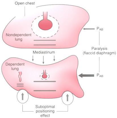
Figure 49-13
Schematic summary of ventilation-perfusion relationships
in an anesthetized patient in the lateral decubitus position who has an open chest
and is paralyzed and suboptimally positioned. The nondependent lung is well ventilated
(as indicated by the large dashed lines) but poorly
perfused (small perfusion vessel); the dependent lung is poorly ventilated (small
dashed lines) but well perfused (large perfusion vessel). In addition,
an atelectatic shunt compartment (indicated on the left side of the lower lung) may
also develop in the dependent lung because of the circumferential compression of
this lung. (See the text for a detailed explanation.) PAB
, pressure of
the abdominal contents. (Modified from Benumof JL: Anesthesia for Thoracic
Surgery. Philadelphia, WB Saunders, 1987.)