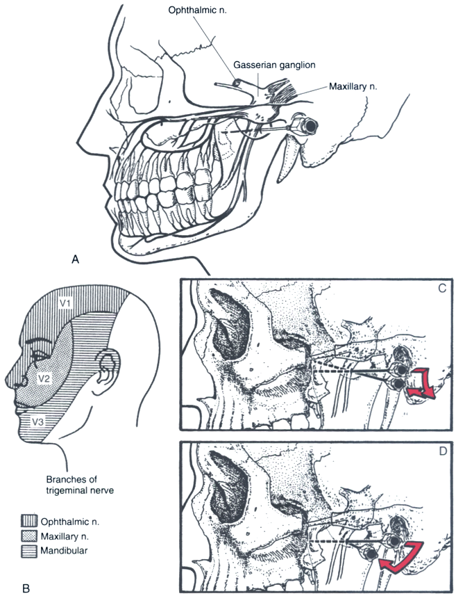
Figure 44-20
Anatomic landmarks and method of needle placement for
maxillary and mandibular nerve blocks. A, The ophthalmic,
maxillary, and mandibular nerves provide sensation to the eye and forehead, midface
and upper jaw, and lower jaw, respectively. B, The
mandibular and maxillary nerves can be blocked through the same needle entry site.
C, A needle is inserted at the inferior edge of the
coronoid notch perpendicular to the skin entry site, and after contacting the lateral
pterygoid plate, it is withdrawn and redirected anteriorly and superiorly to walk
off the plate (arrows). It is advanced approximately
0.5 cm into the pterygopalatine fossa. D, The mandibular
nerve is blocked through the same entry site as the maxillary nerve and by the same
method, except that the needle is withdrawn and redirected to walk off the posterior
border of the pterygoid plate (arrow). When it is
advanced to elicit paresthesia, the needle should not be inserted more than 0.5 cm
past the plate.

