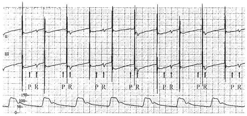
Figure 35-7
Improper placement of a transesophageal pacemaker. The
top recording is electrocardiographic (ECG) lead
II, the middle recording is ECG lead III, and the
bottom recording is the invasive arterial pressure
waveform. This 72-year-old man developed a sinus bradycardia with evidence of tissue
underperfusion. A transesophageal pacemaker was placed, and the large electrocardiographic
artifacts at 75 beats/min were misinterpreted as ventricular systoles (i.e., capture).
They were the pacing stimuli as represented on the ECG monitor. This patient has
a sinus rate of 50 beats/min with a first-degree atrioventricular block (PR interval
of 280 msec). The patient's native atrial (P) and ventricular (R) depolarizations
are identified. The arterial pressure waveform confirms pacing noncapture.