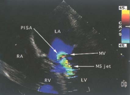
Figure 33-3
Color Doppler imaging of severe mitral stenosis. This
four-chamber echocardiogram reveals a thickened and narrowed mitral valve (MV) indicative
of mitral stenosis (MS). Color Doppler demonstrates (1) acceleration of blood flow
into the stenotic valve (a light blue semicircular area immediately above the valve
called "PISA"—proximal isovelocity surface area), (2) a narrow color jet across
the valve itself, and (3) a 1 by 4-cm color jet extending from the undersurface of
the valve into the left ventricle. LA, left atrium; LV, left ventricle; RA, right
atrium; RV, right ventricle. (From Cahalan MK: Intraoperative Transesophageal
Echocardiography. An Interactive Text and Atlas. New York, Churchill Livingstone,
1997.)

