 |
 |
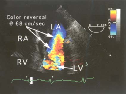
Figure 33-1
"Normal" color Doppler aliasing. In this echocardiogram,
"normal" color Doppler aliasing is seen because laminar flow of blood through the
mitral valve and into the left ventricle exceeds the Nyquist limit (68 cm/sec in
this example—see the color reference icon at the upper right of the figure),
thereby resulting in reversal of the color coding of flow direction. Notice that
this color reversal occurs across fairly broad, regular areas and not in a random
or point-by-point fashion as occurs with turbulent flow (always abnormal). In this
example, follow the blue flow from high in the left atrium as it accelerates into
the mitral orifice, and notice how color Doppler depicts the increasing flow velocities:
the blue color becomes lighter and lighter until the Nyquist limit is reached.
Then, color reversal occurs, with light blue becoming yellow. Just at that reversal
point, the velocity equals the Nyquist limit, in this example 68 cm/sec. Subsequent
reversals may occur at that limit or at multiples of that limit. LA, left atrium;
LV, left ventricle; RA, right atrium; RV, right ventricle. (From Cahalan
MK: Intraoperative Transesophageal Echocardiography. An Interactive Text and Atlas.
New York, Churchill Livingstone, 1997.)

 |
