 |
|
|
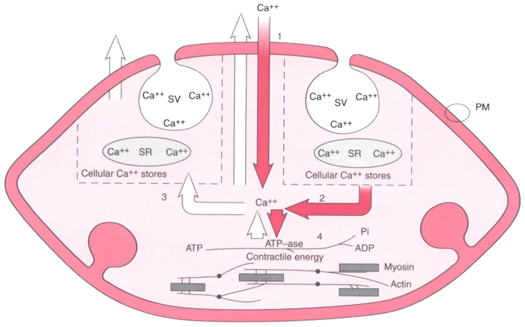
Figure 27-23
Schematic drawing of a smooth muscle cell showing calcium
flux and possible sites of interference by halothane and nifedipine. The concentration
of calcium (Ca2+
) in the cytoplasm increases (red
arrows) because of entry through the plasma membrane (PM) and release
from surface vesicles (SV) or the sacroplasmic reticulum (SR). When the concentration
of cytoplasmic Ca2+
is sufficiently high, adenosine triphosphate (ATP)
is activated. Splitting of ATP by adenosine triphosphatase (ATPase) into phosphatidylinositol
(Pi) and adenosine diphosphate (ADP) provides the interaction and contraction of
actin filaments and myosin particles constituting muscle fibers. The concentration
of cytoplasmic Ca2+
decreases (white arrows)
with the return of Ca2+
to cellular stores and the extracellular transport
of Ca2+
. Both halothane and nifedipine probably (1) inhibit the entry
of Ca2+
and (2) may also interfere with cytoplasmic Ca2+
flux
by reducing the release of Ca2+
by the SR, by (3) reducing storage and
reuptake, or by (4) blocking ATPase or the contractile mechanism (or both). (Redrawn
from Tosone SR, Reves JG, Kissin I, et al: Hemodynamic responses to nifedipine in
dogs anesthetized with halothane. Anesth Analg 62:903, 1983.)
|
|