 |
 |
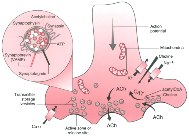
Figure 22-2
The working of a chemical synapse, the motor nerve ending,
including some of the apparatus for transmitter synthesis. The large, intracellular
structures are mitochondria. Acetylcholine, synthesized from choline and acetate
by acetylcoenzyme A, is transported into coated vesicles, which are moved to release
sites. A presynaptic action potential, which triggers calcium influx through specialized
proteins (Ca2+
channels), causes the vesicles to fuse with the membrane
and discharge transmitter. Membrane from the vesicle is retracted from the nerve
membrane and recycled. Each vesicle can undergo various degrees of release of contents—from
incomplete to complete. The transmitter is inactivated by diffusion, catabolism,
or reuptake. The inset provides a magnified view
of a synaptic vesicle. Quanta of acetylcholine together with ATP are stored in the
vesicle and covered by vesicle membrane proteins. Synaptophysin is a vesicle membrane
component glycoprotein. Synaptotagmin is the vesicle's calcium sensor. Phosphorylation
of another membrane protein, synapsin, facilitates vesicular trafficking to the release
site. Synaptobrevin (VAMP) is a SNARE protein involved in attaching the vesicle
to the release site (see Fig. 22-3
).
ACh, acetylcholine, acetyl CoA, acetyl coenzyme A; CAT, choline acetyltransferase.

 |
