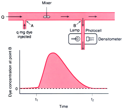
Figure 18-8
Illustration demonstrating the principle of determination
of cardiac output by using the indicator dilution technique. This model assumes
that there is no recirculation. A known amount of dye (q) is injected at point A
into a stream flowing at  (mL/min). A mixed sample of the fluid flowing past
point B is withdrawn at a constant rate through a densitometer. The change in dye
concentration over time is depicted in a curve.
Flow may be measured by dividing the amount of indicator injected upstream by the
area under the downstream concentration curve. (From Berne RM, Levy MN:
The cardiac pump. In Cardiovascular Physiology,
8th ed. St Louis, CV Mosby, 2001, pp 55–82.)
(mL/min). A mixed sample of the fluid flowing past
point B is withdrawn at a constant rate through a densitometer. The change in dye
concentration over time is depicted in a curve.
Flow may be measured by dividing the amount of indicator injected upstream by the
area under the downstream concentration curve. (From Berne RM, Levy MN:
The cardiac pump. In Cardiovascular Physiology,
8th ed. St Louis, CV Mosby, 2001, pp 55–82.)