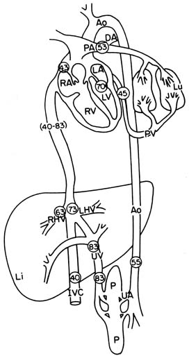
Figure 58-3
The fetal circulation demonstrating the major blood flow
patterns and oxygen saturation values (circled numbers
highlight percent saturation). Ao, aorta; DA, ductus arteriosus; DV, ductus venosus;
IVC, inferior vena cava; Li, liver; Lu, lung; P, placenta; PA, pulmonary artery;
PV, pulmonary vein; RA and LA, right and left atria; RHV and LHV, right and left
hepatic veins; RV and LV, right and left ventricles; SVC, superior vena cava; UA,
umbilical artery; UV, umbilical vein. (From Birnbach DJ, Gatt SP, Datta
S [eds]: Textbook of Obstetric Anesthesia. New York, Churchill Livingstone, 2000,
p 51.)

