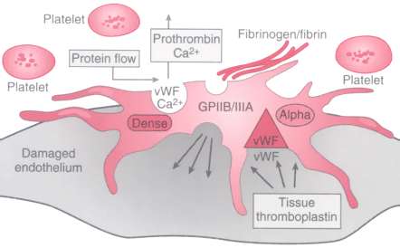
Figure 50-42
Schematic diagram of an activated platelet. Normal platelets
in their resting state are discoid in appearance and very pliable; therefore, they
are able to roll and flow through the vasculature. When activated, they change their
shape to spiculated, and once adherent to a surface, they spread in an ameboid shape.
Once activated, they release the contents of their granules and express a number
of glycoprotein binding sites, of which GPIIB/IIA is an example. GPIIB/IIIA is the
binding site for fibrinogen and fibrin and is the most prolific cellular ligand known.
vWF, von Willebrand factor. (Redrawn from Miller RD, Lichtor JL [eds]:
Atlas of Anesthesia, vol III, Preoperative Preparation and Intraoperative Monitoring.
Philadelphia, Churchill Livingstone, 1997, page 15.4.)