|
|
|
|
|
|
|
|
|
|
|
|
|
|
|
The three-electrode system uses only three electrodes to record the ECG. In such a system, the ECG is observed along one bipolar lead between two of the electrodes while the third electrode serves as a ground. A selector switch allows the user to alter the designation of the electrodes. Three electrocardiographic leads can be examined in sequence without changing the location of the electrodes. Although the three-electrode system has the advantage of simplicity, its use is limited in myocardial ischemia because it provides a narrow picture of myocardial electrical activity.
Numerous modifications of the standard bipolar limb lead system have been developed. Some of these are displayed
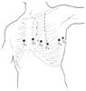 Figure 34-5
Anatomic location of the precordial unipolar leads V1
through V6
. (From Thys DM, Kaplan JA: The ECG in Anesthesia
and Critical Care. New York, Churchill Livingstone, 1987.)
Figure 34-5
Anatomic location of the precordial unipolar leads V1
through V6
. (From Thys DM, Kaplan JA: The ECG in Anesthesia
and Critical Care. New York, Churchill Livingstone, 1987.)
The central subclavicular (CS5 ) lead is particularly well suited for the detection of anterior myocardial wall ischemia. The right arm (RA) electrode is placed under the right clavicle, the left arm (LA) electrode is placed in the V5 position, and the left leg electrode is in its usual position to serve as a ground. Lead I is selected for detection of anterior wall ischemia, and lead II can be selected for monitoring inferior wall ischemia or for the detection of arrhythmias. If a unipolar precordial electrode is unavailable, the CS5 bipolar lead is the best and easiest alternative to a true V5 lead for monitoring myocardial ischemia.
The central back (CB5 ) lead is good for the detection of ischemia and supraventricular arrhythmias as demonstrated in a study comparing CB5 and V5 in patients with closed and open chests.[6] The P wave was 90% larger than in lead V5 , whereas a good correlation between ventricular deflections of CB5 and V5 leads was observed. CB5 is obtained by placing the RA electrode over the center of the right scapula and the LA electrode in the V5 position. The lead selector switch should be on lead I. The CB5 lead may be useful in patients with ischemic heart disease who are susceptible to the development of arrhythmias during the perioperative period.
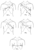 Figure 34-6
Modified bipolar standard limb lead system: MCL1
,
CS5
, CM5
, CB5
, CC5
. (From Thys
DM, Kaplan JA: The ECG in Anesthesia and Critical Care. New York, Churchill Livingstone,
1987.)
Figure 34-6
Modified bipolar standard limb lead system: MCL1
,
CS5
, CM5
, CB5
, CC5
. (From Thys
DM, Kaplan JA: The ECG in Anesthesia and Critical Care. New York, Churchill Livingstone,
1987.)
When modified bipolar limb leads are used, monitoring personnel should be aware that in certain aspects these leads differ significantly from true unipolar precordial leads. The modified precordial leads usually show greater R-wave amplitude than standard precordial leads, and this feature can result in amplification of the ST-segment response.
The criteria for diagnosing myocardial ischemia (discussed later) may need to be adjusted when modified bipolar leads are used. It has been shown during exercise stress testing that normalization of the degree of ST-segment depression to the height of the R wave increases the sensitivity and specificity of ECG for the recognition of myocardial ischemia.[7] In an intra-operative study, Mark and coworkers[8] observed that placement of a Canadian sternal retractor during cardiac surgery was associated with a reduction in V5 R-wave amplitude from 15 ± 1 to 10 ± 1 mm. Simultaneously, V5 S-wave amplitude and absolute ST-segment deviation were reduced from 3.5 ± 0.4 to 1.7 ± 0.3 mm and from 0.50 ± 0.04 to 0.39 ± 0.05 mm, respectively ( Table 34-2 ). The researchers concluded that their results support the proposal that inclusion of an R-wave gain factor may improve perioperative electrocardiographic monitoring.
The use of five electrodes allows the recording of the six standard limb leads (I, II, III, aVR, aVL, aVF), as well as one precordial unipolar lead. Generally, the unipolar lead is placed in the V5 position, which is along the anterior axillary line in the fifth intercostal space ( Fig. 34-7 ). With the addition of only two electrodes to the electrocardiographic system, up to seven different leads can be monitored simultaneously. This allows monitoring of several areas of the myocardium for ischemia or establishing a differential diagnosis between atrial and ventricular arrhythmias.
In 1976, Kaplan and King[9] suggested monitoring lead V5 as the best choice for the detection of intraoperative ischemia. Using a single lead in high-risk patients undergoing noncardiac surgery, London and colleagues[10] demonstrated that the greatest sensitivity was obtained with lead V5 (75%), followed by lead V4 (61%). Combining leads V4 and V5 increased the sensitivity to 90%, whereas with the standard lead II and V5 combination, the sensitivity was only 80%. London and colleagues also suggested that if three leads (II, V4 , and V5 ) could be examined simultaneously, the sensitivity would rise to 98%. Most modern electrocardiographic monitors, however, do not readily allow the simultaneous display of more than one precordial lead. Some investigators
| Variable | Period | Mean ± SEM | n Increase | n Decrease | X2 | P |
|---|---|---|---|---|---|---|
| V5 RWA | Baseline | 15.1 ± 0.8 | — | — | — | — |
|
|
Retractor | 9.9 ± 0.7 | 9 | 69 | 44.6 | 0.001 |
| V5 SWA | Baseline | 3.5 ± 0.4 | — | — | — | — |
|
|
Retractor | 1.7 ± 0.3 | 3 | 59 | 48.8 | 0.001 |
| V5 ST-ABS | Baseline | 0.50 ± 0.04 | — | — | — | — |
|
|
Retractor | 0.40 ± 0.05 | 20 | 51 | 12.68 | 0.001 |
| n, number of patients showing the indicated change; RWA, R-wave amplitude; ST-ABS, absolute value of ST displacement; SWA, S-wave amplitude; X2 , comparison between n increase and n decrease. | ||||||
| Adapted from Mark JB, Chien GL, Steinbrook RA, et al: Electrocardiographic R-wave changes during cardiac surgery. Anesth Analg 74:26, 1992. | ||||||
Great individual variability in the placement of precordial leads is a problem that has been recognized. In an attempt to reduce the error, Herman and coworkers[12] studied a device that facilitates precordial lead placement. The ECG obtained after device-guided electrode placement showed variations from those obtained without it in 60% of patients. Significant Q-wave appearance and disappearance or significant ST-segment elevation and depression, or both, occurred in 19% of patients.
The electrical potentials of the heart can be measured from a surface ECG and from body cavities adjacent to the heart (i.e., esophagus or trachea) or from within the heart itself.
The concept of esophageal ECG is not new; numerous studies have demonstrated the usefulness of this approach in
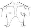 Figure 34-7
Multiple-lead electrocardiographic system, consisting
of four extremity electrodes and the V5
lead. LA, left arm; LL, left
leg; RA, right arm; RL, right leg. (From Narang J, Thys DM: Electrocardiographic
monitoring. In Ehrenwerth J, Eisenkraft JB [eds]:
Anesthesia Equipment: Principles and Applications. St. Louis, Mosby-Year Book,
1992, p 284.)
Figure 34-7
Multiple-lead electrocardiographic system, consisting
of four extremity electrodes and the V5
lead. LA, left arm; LL, left
leg; RA, right arm; RL, right leg. (From Narang J, Thys DM: Electrocardiographic
monitoring. In Ehrenwerth J, Eisenkraft JB [eds]:
Anesthesia Equipment: Principles and Applications. St. Louis, Mosby-Year Book,
1992, p 284.)
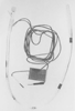 Figure 34-8
In the cardioesophagoscope, the esophageal leads are
made of plastic. The electrocardiographic (ECG) wires are connected to the right
and left arms, with lead I selected on the electrocardiographic monitor. (Adapted
from Narang J, Thys DM: Electrocardiographic monitoring. In
Ehrenwerth J, Eisenkraft JB [eds]: Anesthesia Equipment: Principles and Applications.
St. Louis, Mosby-Year Book, 1992, p 284.)
Figure 34-8
In the cardioesophagoscope, the esophageal leads are
made of plastic. The electrocardiographic (ECG) wires are connected to the right
and left arms, with lead I selected on the electrocardiographic monitor. (Adapted
from Narang J, Thys DM: Electrocardiographic monitoring. In
Ehrenwerth J, Eisenkraft JB [eds]: Anesthesia Equipment: Principles and Applications.
St. Louis, Mosby-Year Book, 1992, p 284.)
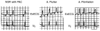 Figure 34-9
Enhanced detection of atrial electrical activity using
an esophageal electrocardiogram (EsECG). The EsECG demonstrates arrhythmia progression
from normal sinus rhythm (NSR), with one premature contraction (PAC), to atrial flutter
(A flutter) and atrial fibrillation (A fibrillation). Lack of information about
atrial activity from lead V5
made definitive diagnosis impossible. (Adapted
from Thys DM, Kaplan JA: The ECG in Anesthesia and Critical Care. New York, Churchill
Livingstone, 1987.)
Figure 34-9
Enhanced detection of atrial electrical activity using
an esophageal electrocardiogram (EsECG). The EsECG demonstrates arrhythmia progression
from normal sinus rhythm (NSR), with one premature contraction (PAC), to atrial flutter
(A flutter) and atrial fibrillation (A fibrillation). Lack of information about
atrial activity from lead V5
made definitive diagnosis impossible. (Adapted
from Thys DM, Kaplan JA: The ECG in Anesthesia and Critical Care. New York, Churchill
Livingstone, 1987.)
Several investigators have described devices that allow electrocardiographic
recording from the esophagus and pacing of the heart using the same device.[14]
Esophageal electrodes have been found particularly useful in patients
|
|
|
Correct Diagnosis from Single Leads | ||
|---|---|---|---|---|
| Arrhythmia | No. | V5 | II | Esophageal |
| Sinus bradycardia | 4 | 4 | 4 | 4 |
| Sinus tachycardia | 1 | 1 | 1 | 1 |
| First-degree heart block | 1 | 0 | 0 | 1 |
| Second-degree heart block | 2 | 0 | 0 | 2 |
| Third-degree heart block | 4 | 0 | 1 | 4 |
| Frequent premature ventricular contractions | 2 | 2 | 2 | 2 |
| Frequent premature atrial contractions | 5 | 2 | 3 | 5 |
| Atrial flutter | 2 | 0 | 0 | 2 |
| Atrial fibrillation | 3 | 2 | 2 | 3 |
| Paroxysmal atrial tachycardia | 1 | 0 | 0 | 1 |
| Nodal rhythm | 1 | 0 | 0 | 1 |
| Percent correct |
|
42.3% | 53.8% | 100% |
| Adapted from Kates RA, Zaidan JR, Kaplan JA: Esophageal lead for intraoperative electrocardiographic monitoring. Anesth Analg 61:781, 1982. | ||||
For many years, long saline-filled central venous catheters have been used to record an intracardiac ECG. More recently, Chatterjee and colleagues [17] described the use of a modified balloon-tipped flotation catheter for recording an intracavitary ECG ( Fig. 34-11 ). The multipurpose pulmonary artery catheter that is available has all the features of a standard pulmonary artery catheter. Three atrial and two ventricular electrodes have been incorporated into the catheter. These electrodes permit recording of an intracavitary ECG and the establishment of atrial or atrioventricular pacing. The diagnostic capabilities with this catheter are great because atrial, ventricular, or atrioventricular nodal arrhythmias and conduction blocks can be demonstrated. The large voltages obtained from the intracardiac electrodes are relatively insensitive to electrocautery
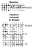 Figure 34-10
Tracings of the esophageal electrocardiogram (EsECG)
and of leads II and V5
indicate posterior ischemia. (Adapted
from Kates RA, Zaidan JR, Kaplan JA: Esophageal lead for intraoperative electrocardiographic
monitoring. Anesth Analg 61:781, 1982.)
Figure 34-10
Tracings of the esophageal electrocardiogram (EsECG)
and of leads II and V5
indicate posterior ischemia. (Adapted
from Kates RA, Zaidan JR, Kaplan JA: Esophageal lead for intraoperative electrocardiographic
monitoring. Anesth Analg 61:781, 1982.)
The endotracheal ECG allows monitoring of the ECG when it is impractical or impossible to monitor the surface ECG. The endotracheal ECG consists of a standard
 Figure 34-11
The multipurpose pacing pulmonary artery catheter. Three
atrial and two ventricular electrodes can be seen. (From Narang J, Thys
DM: Electrocardiographic monitoring. In Ehrenwerth
J, Eisenkraft JB [eds]: Anesthesia Equipment: Principles and Applications. St.
Louis, Mosby-Year Book, 1992, p 284.)
Figure 34-11
The multipurpose pacing pulmonary artery catheter. Three
atrial and two ventricular electrodes can be seen. (From Narang J, Thys
DM: Electrocardiographic monitoring. In Ehrenwerth
J, Eisenkraft JB [eds]: Anesthesia Equipment: Principles and Applications. St.
Louis, Mosby-Year Book, 1992, p 284.)
The clinical use during angioplasty of a coronary guide wire for the recording of intracoronary ECG was first reported in 1985.[20] The major advantage was perceived to be a greater detection of acute ischemia than with surface ECG. In a study of 300 consecutive patients, intracoronary ECG detected ST-segment changes in 83% of lesions, compared with 67% on surface ECG.[21] Maeda and coworkers[22] observed a more rapid shortening of the QT interval with intracoronary ECG than with surface ECG after angioplasty balloon inflation.
|
|
|
|
|
|
|
|
|
|
|
|
|