Passive Electrical Examination (Electrocardiograph,
Electroencephalograph)
Electrocardiograph
Now that some of the basics of electricity have been described,
we can discuss the electrocardiograph (ECG) and the electroencephalograph (EEG),
where the sources of EMF are the heart and the brain. Electrical potentials on biologic
surfaces are too small to observe directly and must be amplified and processed before
display. ECG potentials on the skin are in the 1-mV range, and EEG potentials are
near 0.1 mV.
Figure 30-31
illustrates why electrical potentials on biologic surfaces are so small. The heart
generates an electrical
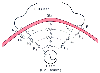 Figure 30-31
Why electrocardiographic potentials are so small. Multiple
resistances and capacitances in the body decrease the potential and distort the waveform
before the electromotive force reaches the surface.
Figure 30-31
Why electrocardiographic potentials are so small. Multiple
resistances and capacitances in the body decrease the potential and distort the waveform
before the electromotive force reaches the surface.
signal as a result of the synchronous depolarization and repolarization of multiple
cells. The electrical potentials generated by the heart are measured by two skin
electrodes, A and B. As the figure shows, there are multiple effective resistances
and capacitances in the tissues between the EMF source and the measuring electrodes.
These impedances lower the magnitude of the voltage signal at the skin. The "shunt"
resistors R3
, R4
, and R5
combined with the "series"
resistors R1
, R2
, and R3
form what is called a voltage
divider network. Lower values of shunt resistance or higher values of
series resistance result in smaller voltages at the skin. The capacitance of the
skin (Cs
) also acts to attenuate the low-frequency components and distort
the waveform. Skin resistance, which may be a megaohm (106
ohms) for
dry skin, can be reduced to a few hundred ohms by conductive gels.
If a DC voltage is applied between two body surface electrodes,
current flows through the tissues between them. Although the electrical current
in metals consists entirely of electron flow, in tissues, both positive and negative
ions migrate. Negative ions tend to accumulate at the positive electrode (the anode),
and positive ions accumulate at the negative electrode (the cathode). This collection
of anions and cations near each electrode creates its own EMF, and this force opposes
the EMF that set up the original current. The current therefore decreases, so the
effective impedance between the electrodes increases. This phenomenon, called polarization
of electrodes, has two harmful effects. First, the increased impedance
from polarization can attenuate the ECG signal for several seconds after defibrillation
or DC cardioversion. Such attenuation could be misinterpreted as a lack of electrical
activity and result in inappropriate administration of a second shock. The second
consequence of prolonged application of DC voltage is accumulation of a local concentration
of toxic ions near electrode sites, a condition that can cause burns or tissue necrosis.
A partial solution to the problem is the use of a nonpolarizable electrode, such
as a silver and silver chloride combination. This electrode can act as a source
of, or "sink," for both anions and cations, thereby minimizing the accumulation of
ions. Most disposable ECG electrodes now use such materials. Even these electrodes,
however, are nonpolarizable for only a limited time, and the application of prolonged
DC voltage between any tissue electrodes must be avoided.
Electroencephalograph
Similar problems complicate measurements of the EEG, but in this
case, the signal is only one tenth the amplitude: 100 µV versus 1 mV for the
ECG. The spontaneous surface EEG provides eight or more channels of amplitude (surface
voltage)-versus-time data, which is of limited use for monitoring in the operating
room. For rapid interpretation and diagnosis, the amplitude-versus-time data are
usually transformed into plots of amplitude versus frequency. This process of power
spectral analysis has been discussed in the section on signal processing. The EEG
power spectrum facilitates rapid diagnosis of hemisphere asymmetries and changes
in frequency content that accompany either deep anesthesia or cerebral hypoxia.
"Bispectral density" is a newer method of analyzing the EEG that determines levels
of correlation between various frequency components in the power spectrum. This
type of analysis may provide improved determination of the depth of anesthesia in
some settings.