Intravascular Pressures
The first measurement of the blood pressure was made by Stephen
Hales (1677–1761) ( Fig. 1-3A
),
the curate of Middlesex, England, who between sermons occupied himself with experiments
on the mechanics of the circulation. He[25]
described
one of his experiments performed (1733) on an "old mare who was to be killed, as
being unfit for service":
I fixed a brass pipe to the carotid artery of a mare ... the blood rose in the
tube till it reached to nine feet six inches in height. I then took away the tube
from the artery and let out sixty cubic inches of blood, and then replaced the tube
to see how high the blood would rise after each evacuation; this was repeated several
times until the mare expired. In the three horses, death occurred when the height
of the blood in the tube was about two feet.
Hales also discovered that the resistance of a vascular bed could
change by mixing alcohol in the blood, which he observed could account for changes
in blood pressure brought about by diverse ingested agents. In 1828, Jean L. Poiseuille
[26]
(1799–1869) repeated these experiments
and devised a hemomanometer that used mercury instead of the long blood-filled tubes
used by Hales. Poiseuille also showed that the blood pressure varied with respiration.
In 1854, Karl Vierrordt[27]
(1818–1884)
invented a sphygmograph, acting on the principle that indirect estimation of blood
pressure could be accomplished by measuring the counterpressure necessary to obliterate
the arterial pulsation. Scipione Riva Rocci's (1863–1937) sphygmomanometer,
described in 1896,[28]
used the same principle but
used a rubber cuff that occluded a major
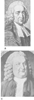 Figure 1-3
Two scientists of the Enlightenment era. A,
Stephen Hales (1677–1761): detail of an oil painting by T. Hudson, 1759, in
the National Portrait Gallery, London. Hales was educated at Cambridge University,
England and was ordained as a minister in 1703. He spent his career as minister
to the parish of Teddington, England. His rudimentary studies on the gas produced
by mixing Walton pyrites (i.e., ferric disulfide) and spirit of nitre (i.e., nitric
acid) was the spark that prompted Priestley to pursue his studies on nitric oxide,
which led to the discovery of nitrous oxide in 1773. Hales was the first to measure
blood pressure and cardiac output. He also developed ventilators that brought fresh
air into prisons and granaries. B, Albrecht von Haller
(1708–1777): detail of an engraving by Ambroise Tardieu. Haller was born
in Bern, Switzerland. He served as professor of medicine and surgery at the University
of Göttingen, Germany, where he began his encyclopedic work, Physiological
Elements of the Human Body, published in eight volumes between 1757 and
1766. His demonstration that "irritability" was a property of muscle and "sensitivity"
was a property of nerves was derived from nearly 600 experiments on live animals.
He returned to Bern in 1753, and while there he published a catalog of the scientific
literature containing 52,000 references. (Portraits courtesy of the National
Library of Medicine, Bethesda, MD.)
Figure 1-3
Two scientists of the Enlightenment era. A,
Stephen Hales (1677–1761): detail of an oil painting by T. Hudson, 1759, in
the National Portrait Gallery, London. Hales was educated at Cambridge University,
England and was ordained as a minister in 1703. He spent his career as minister
to the parish of Teddington, England. His rudimentary studies on the gas produced
by mixing Walton pyrites (i.e., ferric disulfide) and spirit of nitre (i.e., nitric
acid) was the spark that prompted Priestley to pursue his studies on nitric oxide,
which led to the discovery of nitrous oxide in 1773. Hales was the first to measure
blood pressure and cardiac output. He also developed ventilators that brought fresh
air into prisons and granaries. B, Albrecht von Haller
(1708–1777): detail of an engraving by Ambroise Tardieu. Haller was born
in Bern, Switzerland. He served as professor of medicine and surgery at the University
of Göttingen, Germany, where he began his encyclopedic work, Physiological
Elements of the Human Body, published in eight volumes between 1757 and
1766. His demonstration that "irritability" was a property of muscle and "sensitivity"
was a property of nerves was derived from nearly 600 experiments on live animals.
He returned to Bern in 1753, and while there he published a catalog of the scientific
literature containing 52,000 references. (Portraits courtesy of the National
Library of Medicine, Bethesda, MD.)
arterial vessel and then slowly deflated. In 1905, Nikolai Korotkov[29]
(1874–1920) described the sounds produced during auscultation over a distal
portion of the artery as the cuff was deflated. The Korotkov sounds resulted in
more accurate determinations of systolic and diastolic blood pressures. Oscillometric
blood pressure measurements relied on a cuff that sensed the changes in arterial
pulsations and was described by H. von Recklinghausen in 1931.[30]
Automatic blood pressure devices based on the oscillometric method were developed
in the 1970s and have become the standard noninvasive measures of arterial pressure
in most hospitals.
The past 50 years have seen a gradual return to direct measurements
of arterial blood pressure, which are in principle much the same method used by Stephen
Hales nearly 250 years ago. However, the Poiseuille method of using mercury-filled
glass tubes was found to be totally inadequate for recording accurate pressures in
a dynamic system. In 1876, Herbert Tomlinson[31]
introduced the principle of the strain gauge: resistance in a wire increases when
it is stretched. It took 70 years after this principle was described before a strain
gauge was used to measure blood pressure. In 1947, at the Mayo Clinic in Rochester,
Minnesota, E. H. Lambert and E. H. Wood[32]
first
reported the use of a strain gauge to measure blood pressure continuously in human
subjects exposed to acceleration forces. Cannulas placed directly into vessels were
first described in 1949,[33]
and since then, direct
measurements of blood pressure expanded gradually into the operating room and intensive
care units.
Venous pressures were of less interest to anesthesiologists until
convenient methods for placing cannulas into central vascular structures were described
50 years ago by Sven Seldinger.[34]
Werner Forssman
(1904–1979), a urologist, described the methods of central venous access and
right heart catheterization in humans in 1929,[35]
originally experimenting on himself, and was awarded the Nobel Prize in Physiology
and Medicine for his work on venous pressures in 1956. The introduction of plastic
catheters[36]
gradually made it possible to measure
central pressures in the clinical setting. Although arm and femoral veins were used
initially, subclavian and internal jugular vein cannulation eventually replaced the
peripheral sites. Pulmonary artery catheterization with a balloon-tipped, flow-directed
catheter was described in 1970[37]
and has been
used extensively since then by anesthesiologists to measure cardiac outputs using
the Fick[38]
principle and pulmonary wedge pressures.
The pulmonary artery catheter also allowed the clinician to use the well-known pressure-volume
relationships of the heart described by Ernest H. Starling (1866–1927) in 1918
to maximize cardiac outputs and oxygen delivery to the tissues.
Transesophageal echocardiography (TEE) was described in 1976[39]
and used in anesthesia practice a few years later. One of the original probes used
during anesthesia was fashioned from an esophageal stethoscope combined with an M-mode
echocardiographic probe and was used to calculate cardiac output and ejection fraction
in a 65-year-old woman undergoing mitral valve repair.[40]
An improved electronic phased-array transducer was initially applied at the University
of California, San Francisco for
monitoring regional myocardial function in high-risk surgical patients.[41]
Biplane TEE and color flow mapping were introduced in the 1980s and resulted in
an explosive growth in TEE applications.[42]
With
experience and training in TEE, the anesthesiologist can quickly evaluate filling
pressures of the heart as well as obtain measures of myocardial contractility and
valvular function. TEE has become a routine monitor for certain surgical procedures.