|
|
|
|
|
|
|
|
|
|
|
|
|
|
|
Laryngeal trauma has no standard features (see Chapter 63 ).[126] [127] [128] [129] Internal damage may be extensive with no external signs. Stridor and cyanosis may not be present. These injuries most often result from high-speed blunt trauma. Velocity-deceleration accidents can produce severe closed neck injury to the trachea and larynx. Tracheal damage can also be caused by a burst injury that produces tears, dislocation of arytenoid cartilage, and disruption of the cricothyroid joint. More obvious injuries such as a fractured femur can divert the physician's attention from the more subtle signs of airway problems such as hoarseness, weak voice, or tracheal tugging.
Signs of airway problems may be delayed and include stridor, wheezing, coughing, hemoptysis, retraction of the airway, change in voice, and difficulty speaking or swallowing. Loss of neck prominences and increasing crepitus indicate torn mucosa in the trachea, pharynx, larynx, or esophagus. The amount of crepitus does not correlate with the size of the tear.
An air leak can dissect into the paratracheal space and produce a pneumothorax. Such leakage increases with straining and the application of positive-pressure ventilation. All open neck injuries should be explored because mortality is as high as 5%. The possibility of a cervical spine fracture should be considered whenever head or neck trauma has occurred.
A review of airway management in cervical spine injury indicated that early neck immobilization followed by orotracheal intubation with in-line stabilization had the lowest incidence of adverse events. There is no evidence that nasotracheal intubation is necessarily safer.[130]
The larynx of a young child is positioned high in the neck and is shielded by the mandible. Cartilage is more elastic and flexible in a child and less likely to fracture. Because of weaker connecting membranes, a child is more vulnerable to intercartilage rupture.[126] The airways of young children have a small diameter at the subglottic region of the cricoid cartilage, so they do not tolerate edema of the airway as well.
Airway problems are dynamic. Mild stridor can worsen an air leak and thereby aggravate subcutaneous emphysema
Blind passage of an endotracheal tube can create a false passage through a tear in the mucosa. The trachea should be intubated under direct vision with the use of a laryngoscope, rigid bronchoscope, or fiberoptic laryngoscope. A careful mask induction of anesthesia with minimal application of positive pressure, followed by bronchoscopic examination, may be safe in selected patients. Tracheostomy performed under local anesthesia is often a safer approach.
The most common fractures involve the mandible and midface—Le Fort I, II, and III fractures of the maxilla[129] [131] [132] ( Fig. 65-2 ). All these fractures may be accompanied by some degree of compromised airway. A first priority is to secure the airway by placing the patient in the lateral position, pulling the mandible or maxilla
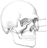 Figure 65-2
Classification of midfacial fractures. Le Fort I: alveolar
fracture. Le Fort II: zygomatic-maxillary complex fracture. Le Fort III: cranial
facial dysostosis with separation of the midface from the skull. (From Schuller
DE, Schleuning AJ: Otolaryngology—Head and Neck Surgery, 8th ed. Mosby-Year
Book, 1994, p 157.)
Figure 65-2
Classification of midfacial fractures. Le Fort I: alveolar
fracture. Le Fort II: zygomatic-maxillary complex fracture. Le Fort III: cranial
facial dysostosis with separation of the midface from the skull. (From Schuller
DE, Schleuning AJ: Otolaryngology—Head and Neck Surgery, 8th ed. Mosby-Year
Book, 1994, p 157.)
Mandibular fractures, if unilateral, are stable, whereas bilateral mandibular fractures are unstable. The posterior fragment may be pulled medially and upward and cause the base of the tongue to obstruct the pharynx. Trismus and lingual hematoma may contribute to respiratory obstruction.[129] If no trismus or mechanical problem exists, routine rapid-sequence induction of anesthesia and tracheal intubation may proceed. Otherwise, induction of anesthesia with an inhaled anesthetic or awake endotracheal intubation is indicated. Although the possibility of interdental fixation may seem to make the use of a nasotracheal tube preferable, the easiest route of intubation should be chosen. Later, during surgery, the oral tube can be removed and a nasal tube inserted. Patients undergoing interdental fixation should be given an antiemetic drug, undergo awake tracheal extubation, and be placed on their side with scissors/wire cutter at the bedside.
Midface fractures, or Le Fort fractures of the maxilla, often present difficult clinical problems. A Le Fort I fracture is a simple horizontal fracture of the lower part of the maxilla that produces only a mobile palate. A Le Fort II fracture, a triangular extension of a Le Fort I fracture, involves two oblique fracture lines along the malomaxillary suture to the floor of the orbit and the base of the tongue. A Le Fort III fracture is a transverse fracture above the malar bone and through the orbits. It is characterized by complete separation of the maxilla from the craniofacial skeleton, epistaxis, and a flat dish-face deformity. Tears in the dura occur in 25% of all Le Fort II and III fractures, along with leakage of cerebrospinal fluid. Orotracheal intubation is necessary with intranasal damage. Attempted passage of a nasogastric or nasotracheal tube may cause bleeding, mucosal dissection, and further damage. These tubes can enter the maxillary antrum, the orbit, the base of the skull, or the cranium.
|
|
|
|
|
|
|
|
|
|
|
|
|