|
|
|
|
|
|
|
|
|
|
|
|
|
|
|
Lung-damaging agents (LDAs) were formally known as choking agents, a term that does not adequately describe the many levels of the lung at which they act. LDAs act at the upper and lower respiratory levels, but their main lethal action is to cause toxic pulmonary edema ( Table 64-8 ). LDAs were the first to be used as chemical weapons during World War I with the use first of chlorine, followed by phosgene. LDAs are a universal hazard in military and civil life because they are also used widely as industrial feedstocks in many chemical engineering processes. The isocyanates are widely used in this respect, and the accidental release of methylisocyanate at Bhopal in India in 1984 caused the largest number of casualties ever recorded after a toxic release in war or peace, with more than 5000 deaths from toxic pulmonary edema.[54] [55] Chemical weapons and HAZMAT sources list many LDAs.[7] [19]
| Military Agents |
| Chlorine |
| Phosgene |
| Perfluoroisobutylene |
| Civilian Agents |
| Phosgene |
| Isocyanates |
| Phenols |
| Glycols and other compounds |
Phosgene has been used as a chemical warfare agent, is an established worldwide industrial hazard, and may be regarded as a potential terrorist threat. It is a model for consideration of other hazards in its class. Phosgene is carbonyl chloride and is a hazard familiar to an older generation of anesthesiologists because it is the product of the reaction of trichloroethylene with soda lime. First synthesized in the 19th century, phosgene has been in industrial use as an intermediary for more than a century but achieved notoriety during World War I, when it was used extensively by both sides from 1915 onward. It caused more than 85% of all deaths from chemical warfare agents.[13] Phosgene is a gas at normal temperatures and is nonpersistent ( Table 64-9 ). It is less water soluble than chlorine, its immediate chemical weapons predecessor in 1915, a feature that results in the gas being taken further down the respiratory tree and into the alveoli.
Phosgene has been known since World War I to have a dual latency
of action, which underlies its essentially dangerous character. Two stages follow
exposure. First, immediately after exposure, there is an intense upper respiratory
irritation with coughing, retching, choking, and chest tightness. These symptoms
are accompanied by eye irritation and lacrimation.[56]
In some cases, death has followed exposure to high concentrations of phosgene without
the
| Properties |
| Chemical name: carbonyl chloride (COCl2 ), chloroformyl chloride |
| Boiling point: 8.2°C |
| Vapor pressure: 1215 at 20°C |
| Vapor density: 3.5 × air |
| Breakdown: slow aqueous hydrolysis to HCl and CO2 |
| Toxic Exposure Levels |
| 1 ppm chronically: chronic lung disease |
| >25 ppm/min: acute lung effects |
| 50–100 ppm/min: initial inflammatory response that may be followed by pulmonary edema |
| >150 ppm/min: clinically significant and life-threatening pulmonary edema |
| Lethal exposure in humans: 800 ppm for 2 minutes |
The lung is a delicately balanced system in which the entire cardiac output passes through the pulmonary circulation arranged as a fine capillary mesh in the interstitial space (i.e., loose organization of collagen, elastin, and various cell types). Because of the fragile nature of structural organization, lung tissue commonly reacts to toxic challenge by producing pulmonary edema, which involves the passage of fluid from the capillaries to the interstitial spaces and then to the interalveolar spaces. Phosgene reacts through covalent attack on many substrate groups, including -NH2 and -SH.[57] Potential cell targets are type I and II pneumocytes and alveolar macrophages. Covalent binding can be seen as the primary attack leading to free radical release. This stage is followed by a secondary attack involving released inflammatory mediators, including prostaglandins, causing vasoconstriction, vasodilatation, and platelet disaggregation; bradykinin, causing increased capillary permeability; 5-hydroxytryptamine (5-HT, serotonin), causing constriction of postcapillary vessels; thromboxane A2 , causing vasoconstriction; and the release of complement-activating enzymes, leading to attraction of leukocytes and leukotriene release.
Patients who have been exposed to phosgene should be removed from the site of exposure as quickly as possible by protected emergency responders. There is usually no requirement for decontamination unless liquid contamination has occurred. There is no specific antidote to phosgene, and treatment is based around use of supportive measures and pharmacologic modification of the effects of the inflammatory mediator cascade.[58] A firm rule for the management of phosgene exposure dating from World War I is complete bed rest and observation for 24 hours. No patient who has had a risk of significant exposure should be discharged from hospital in less than 24 hours unless accompanied by a responsible observer.
Anesthesiologists have a major role to play in the provision of specialized respiratory care at prehospital and hospital levels. Some cases may require early intubation and ventilation, and others require oxygen and supported ventilation in the early stages. The method of provision of emergency ventilation is a matter of interest. Work in physical trauma indicates that the open-lung strategy [59] [60] is of value in the prevention of inflammatory cascades by opening the alveoli and keeping them open by use of the correct level of positive end-expiratory pressure. This technique is based on the hypothesis that repeated opening and closing of alveoli causes kinin release through the action of shear forces in the alveolar walls. This may be an important lesson for the early management of lungs
The use of inhaled and systemic steroids in the treatment of toxic pulmonary edema has been controversial for many years, and Diller,[61] a leading authority on phosgene poisoning, felt in 1985 that the case was unproven. However, there has been renewed experimental interest in the subject of using chlorine, which has produced some encouraging results in pig and rat studies. In a study of 18 pigs subjected to 140 ppm chlorine for
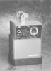 Figure 64-9
The CompPac portable gas-powered emergency and transport
ventilator for use in contaminated environments. (Courtesy of Pneupac Ltd,
Luton, UK.)
Figure 64-9
The CompPac portable gas-powered emergency and transport
ventilator for use in contaminated environments. (Courtesy of Pneupac Ltd,
Luton, UK.)
There have always been species differences in challenges to chemical warfare agents, and results of these studies should be applied with caution to humans. However, in a clinical area for which few therapeutic options are available, the results provide encouragement for further research and for clinical intervention if the need arises.
Kennedy and his group[65] suggested that aminophylline might protect against phosgene-induced pulmonary embolism as a result of its ability to increase cyclic AMP levels. Other compounds, including β-adrenergic agonists, also have this effect and may indicate a new therapeutic direction.
Another promising therapeutic approach is provided by compounds that can increase intracellular levels of reduced glutathione (GSH) as a means of preventing lipid peroxidation-induced pulmonary embolism. The rationale behind this approach is that phosgene reacts with cellular SH groups, reduces the GSH redox state, and increases arachidonic acid mediator production and lipid peroxidation. Sciuto and coworkers[66] studied the effect of N-acetylcysteine (NAC) on anesthetized rabbits exposed to 1500 ppm phosgene. The NAC-treated group had smaller increases in pulmonary wet weight, lower leukotriene levels, and higher glutathione levels. This work suggests that NAC may protect against phosgene-induced pulmonary embolism by maintaining GSH levels and inhibiting production of inflammatory leukotrienes.
The lung-damaging actions of sulfur mustard were previously described. This agent can also cause pulmonary embolism at high ambient temperatures. NAC has been studied in this context and has been reported in a rat study by Anderson and his colleagues[67] to prevent increases in biochemical parameters in lung lavage fluid after exposure to mustard agent. Levels of lactate dehydrogenase and glutamine-oxaloacetic transaminase (i.e., aspartate aminotransferase) did not vary from the control levels, indicating a reduction of cellular injury and transudation. However, NAC was given at the same time as mustard exposure in this study, which is an unlikely timeframe for exposed human casualties. Nevertheless, NAC appears to be a promising line of therapy in an area in which there
|
|
|
|
|
|
|
|
|
|
|
|
|