Late Resuscitation
Table 63-13
summarizes end points for late resuscitation, and Figure
63-10
presents an algorithm for management. Fluid administration is an
integral, mandatory component of late resuscitation. The adequacy of resuscitation
should not be judged by the presence of normal vital signs but by normalization of
organ and tissue perfusion. The role of the anesthesiologist-intensivist is to recognize
the presence of ongoing shock after traumatic hemorrhage and resuscitate the patient
with the appropriate fluid, in the appropriate amount, at the appropriate time.
Late resuscitation begins once bleeding is definitively controlled
by surgery, angiography, or the passage of time. The practitioner's goal at that
time is to rapidly restore normal perfusion to all organ systems while continuing
to support vital functions. Hypoperfusion caused by hemorrhagic shock triggers a
predictable cascade of biochemical events that will cause physiologic derangements
persisting
TABLE 63-13 -- Goals for late resuscitation
*
|
Maintain systolic blood pressure >100 mm Hg |
|
Maintain hematocrit above individual transfusion threshold |
|
Normalize coagulation status |
|
Normalize electrolyte balance |
|
Normalize body temperature |
|
Restore normal urine output |
|
Maximize cardiac output by invasive or noninvasive measurement |
|
Reverse systemic acidosis |
|
Document decrease in lactate to normal range |
*Fluid
administration should be continued until adequate systemic perfusion can be verified.
long after adequate blood flow is restored. The extent of hypoperfusion—the
depth and duration of shock—is highly correlated with the development of subsequent
organ system failure. Unfortunately, traditional vital sign markers such as BP,
heart rate, and urine output have been shown to be insensitive to the adequacy of
resuscitation. Occult hypoperfusion syndrome is common in postoperative trauma patients,
particularly young ones.[97]
This syndrome is characterized
by normal BP maintained by intense systemic vasoconstriction; intravascular volume
is low, cardiac output is low, and organ system ischemia persists. Such patients
are at high risk for MOSF if hypoperfusion is not promptly corrected.
The search for the optimal end point of resuscitation has led
to several different hemodynamic, acid-base, and regional perfusion targets. Table
63-14
summarizes modalities that are available to gauge the adequacy of
resuscitation, along with the shortcomings of each technique. Although the flow
of blood to tissue beds is a determinant of tissue perfusion, pressure should also
be an important consideration. The left ventricular stroke work index is a variable
that accounts for both flow and pressure. Furthermore, left ventricular power output
has been used to quantify left ventricular performance. These indices were compared
with purely flow-derived hemodynamic and oxygen transport variables as markers of
perfusion and outcome in critically injured patients during resuscitation.[98]
A consecutive series of 111 patients were monitored with a volumetric pulmonary
artery catheter during the first 48 hours of resuscitation. The ability to clear
lactate in less than 24 hours and survival were studied. Survivors had significantly
higher stroke work and left ventricular performance than nonsurvivors did. These
variables, in addition to heart rate, were the only ones that were significantly
related to lactate clearance and survival. The higher stroke work and left ventricular
performance
TABLE 63-14 -- Methodologies for assessment of systemic perfusion
|
Technique |
Shortcomings |
|
Vital signs |
Will not indicate occult hypoperfusion |
|
Urine output |
May be confounded by intoxication, diuretic therapy, circadian
variation, or renal injury |
|
Systemic acid-base status |
Confounded by respiratory status |
|
Lactate clearance |
Requires time to obtain laboratory result |
|
Cardiac output |
Requires placement of a pulmonary artery catheter or use of noninvasive
technology |
|
Mixed-venous oxygenation |
Difficult to obtain, but a very accurate marker |
|
Gastric tonometry |
Requires time to equilibrate, subject to artifact |
|
Tissue-specific oxygenation |
Investigational techniques; may not indicate satisfactory systemic
perfusion |
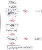 Figure 63-10
Algorithm for the management of late hemorrhagic shock.
HCT, hematocrit; HR, heart rate; PA, pulmonary artery; PT, prothrombin time; SBP,
systolic blood pressure.
Figure 63-10
Algorithm for the management of late hemorrhagic shock.
HCT, hematocrit; HR, heart rate; PA, pulmonary artery; PT, prothrombin time; SBP,
systolic blood pressure.
in survivors were related to better ventricular-arterial coupling and therefore more
efficient cardiac function.
Monitoring resuscitation with clinical variables and monitoring
the adequacy of systemic oxygen delivery with invasive monitors may be supplemented
in the future with an approach that assesses the return of adequate metabolism, respiration,
and oxygen transport in peripheral tissue beds. One minimally invasive technique
that has been proposed is tissue oxygen monitoring (skin, subcutaneous tissue, or
skeletal muscle). Skeletal muscle blood flow decreases early in the course of shock
and is restored late during resuscitation, thus making the skeletal partial pressure
of oxygen a sensitive indicator of low flow.[99]
Tissue hypercapnia has been suggested as a universal indicator
of critically reduced perfusion (also see Chapter
74
). Management of gastric mucosa PCO2
through gastric tonometry has been used in trauma patients as an indicator of restoration
of splanchnic blood flow, and distal gut pH has shown promise as a reliable indicator.
[100]
The esophageal wall has also been demonstrated
to be an appropriate site for tissue PCO2
measurements during hemorrhagic shock,[101]
and
recently, the most proximal area of the gastrointestinal tract, the sublingual mucosa,
has been shown to be a useful site for measurement of PCO2
.
When sublingual PCO2
exceeded a threshold
of 70 mm Hg (normal = 45.2 ± 0.7 mm Hg), its positive
predictive value for the presence of physical signs of circulatory shock was 100%.
[102]
Inadequate tissue perfusion, as indicated
by these specific monitors or by the traditional systemic markers of serum lactate,
base deficit, and decreased pH, must be promptly treated once hemorrhage has been
controlled. The rate at which a shock patient's lactate returns to the normal range
is strongly correlated with outcome: failure to reach the normal range
within 24 hours of a traumatic injury carries a greater risk of organ system failure
and eventual death.[97]
[103]