LOWER EXTREMITY CONDUCTION BLOCKS
Anatomic Considerations
The lower extremity has a more complex nerve supply than the upper
extremity because it depends on two
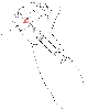 Figure 45-10
Metacarpal (or transthecal) nerve block, showing the
head of the metacarpal located by palpation (1).
Figure 45-10
Metacarpal (or transthecal) nerve block, showing the
head of the metacarpal located by palpation (1).
plexuses: the lumbar (L1 to L4 spinal nerves) and the sacral (L5 to S4) plexuses.
Lumbar Plexus and Lumbar Plexus Nerves
The lumbar plexus supplies the anterior aspect of the limb. It
lies within the substance of the psoas muscle and in a fascial plane called the psoas
compartment, where it can be approached by a posterior route through the
quadratus lumborum muscle. The lumbar plexus contributes four nerves to the lower
extremity: femoral, lateral cutaneous, obturator, and genitofemoral. The psoas
and iliacus muscles share the same aponeuroses, the fascia iliaca. At their emergence
from the psoas muscle, all lumbar plexus nerves run a variable part of their course
just below this fascia. A local anesthetic injected at the inner surface of this
fascia and spreading along it contacts virtually all lumbar plexus nerves if the
injected volume is sufficient. This is the concept sustaining the technique of the
fascia iliaca compartment block.
The femoral nerve is the largest branch of the lumbar plexus.
It emerges from the psoas major muscle in the groove separating the psoas and the
iliacus muscles, passes behind the inguinal ligament, and enters the groin in the
femoral triangle (i.e., Scarpa's triangle). It runs lateral to the femoral vessels
from which it is separated by the lower part of the psoas major muscle. At this
level, it splits into anterior and posterior terminal divisions. Contrary to the
brachial plexus, which is enclosed in the axillary sheath, the femoral nerve remains
outside the sheath surrounding the femoral vessels. Its main division branch, the
saphenous nerve, which is a purely sensory nerve, continues the general direction
of the femoral nerve, lateral to the femoral artery in the adductor canal (i.e.,
Hunter's canal) together with the motor branch supplying the vastus medialis muscle.
At the distal extremity of the adductor magnus muscle, it crosses the femoral vessels
anteriorly, passes behind the sartorius muscle, descends along the medial border
of the tibia, and ends its course on the medial side of the ankle, where it gives
off two terminal branches. In the adductor canal, it gives off twigs to the medial
cutaneous nerve of the thigh and the obturator nerve to form a subsartorial plexus.
It also gives off patellar branches that contribute to the patellar plexus and the
medial cutaneous branches. It supplies the medial part of the leg and the foot.
Sacral Plexus and Sciatic Nerve
The sacral plexus supplies the posterior aspect of the limb.
It lies on the anterior aspect of the piriformis muscle, behind the posterior wall
of the pelvic cavity, and cannot be directly approached by a block needle. It contributes
two nerves to the lower extremity: posterior femoral cutaneous and sciatic. The
sciatic nerve is the largest mixed nerve of the body, and it is formed by two distinct
nerves, the common peroneal nerve and the tibial nerve, which are enclosed in the
same perineural sheath. The sciatic nerve leaves the pelvis through the greater
sciatic foramen and then passes between the greater trochanter of the femur and the
ischial tuberosity. Below the piriformis muscle, it runs in the subgluteal space
within a true aponeurotic canal, in which the pressure is negative and can be detected
by an LOR technique. It is separated from the hip joint by the obturator externus
muscle. In the thigh, the sciatic nerve runs on the adductor magnus, along the lower
border of the femur and toward the popliteal fossa. At the upper part of the popliteal
fossa, the tibial and common peroneal nerves separate. The common peroneal nerve
supplies the lateral aspect of the leg and the dorsum of the foot. The tibial nerve
supplies the dorsal aspect of the leg and the plantar surface of the foot.
 |