Local Anesthesia of the Airway
Clinical Applications
Anesthesia of the airway can be used to facilitate diagnostic
laryngoscopy and bronchoscopy and to allow the comfortable placement of a tracheal
tube in patients whose anatomy dictates an awake endotracheal intubation. Blocks
of the superior laryngeal nerves bilaterally, along with translaryngeal injection
of local anesthetic, provide anesthesia of the airway from the infraglottic area
to the epiglottis. Additional topical application of local anesthetic to the oral
and nasal mucosa, along with appropriate sedation, provides satisfactory analgesia
for endoscopic procedures.
Technique: Superior Laryngeal Nerve
The patient is placed supine with the neck extended. The hyoid
bone is displaced laterally toward the side to be blocked, and a 25-gauge, 2.5-cm
needle is walked off the greater cornu of the hyoid bone inferiorly and is advanced
2 to 3 mm ( Fig. 44-24
).
As the needle passes through the thyrohyoid membrane, a slight loss of resistance
is felt, and 3 mL of local anesthetic solution is injected superficial and deep to
this structure. The block is then repeated on the opposite side. This technique
produces anesthesia from the inferior aspect of the epiglottis to the vocal cords.
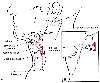 Figure 44-24
A, Anatomic landmarks
and method of needle placement for a superior laryngeal nerve block. B,
The needle is walked off the greater cornu of the hyoid bone inferiorly.
Figure 44-24
A, Anatomic landmarks
and method of needle placement for a superior laryngeal nerve block. B,
The needle is walked off the greater cornu of the hyoid bone inferiorly.
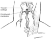 Figure 44-25
Needle placement for a translaryngeal nerve block.
Figure 44-25
Needle placement for a translaryngeal nerve block.
Technique: Translaryngeal Block
The translaryngeal block is simple to perform and results in anesthesia
of the trachea below the vocal cords. However, the injection of local anesthetic
usually stimulates the cough reflex, and this block should be avoided in patients
in whom coughing is undesirable.
With the patient in the supine position, the cricothyroid membrane
is located, and a 20-gauge or smaller, 3- to 5-cm plastic catheter over a needle
is introduced in the midline ( Fig.
44-25
). The inner steel cannula is withdrawn with the plastic catheter
held firmly in place; aspiration of air confirms correct catheter placement. Between
3 and 5 mL of 4% lidocaine solution is injected rapidly, usually
resulting in a vigorous cough, which aids in the spread of the solution within the
trachea.
Technique: Intraoral Approach to Glossopharyngeal
Nerve Block
The glossopharyngeal nerve (i.e., cranial nerve IX) supplies sensation
to the posterior one third of the tongue, the pharynx, and the superior surface of
the epiglottis. It can be blocked intraorally by injecting 5 mL of local anesthetic
into the base of each posterior tonsillar pillar. An angled 22-gauge, 9-cm needle,
which can be formed by bending the distal 1 cm of a spinal needle with its stylet
removed, is employed for this block. Visualization of the posterior pillar is facilitated
by the gentle use of a no. 3 MacIntosh laryngoscope blade after topical anesthetic
has been applied to the tongue. Careful aspiration before injection is mandatory
because of the proximity of the carotid artery.
Side Effects and Complications
The mucosa of the upper airway is well perfused, resulting in
rapid uptake of local anesthetics injected or topically applied in this area. Careful
attention to total drug dosages, close observation of the patient, and compulsive
aspiration before injection diminish the risk of local anesthetic toxicity. Other
problems and complications are rare; however, caution should be employed in the patient
with a full stomach, because these blocks abolish the airway reflexes.