Femoral Nerve Block
The femoral nerve is formed within the psoas major muscle by posterior
divisions of the second, third, and fourth lumbar nerves. It emerges from the lateral
border of the psoas muscle to descend in the groove between the psoas and iliacus
muscles and enters the thigh by passing beneath the inguinal ligament lateral to
the femoral artery. At this point, the nerve divides into multiple terminal
branches, which have been classified as anterior and posterior. The anterior branches
are primarily cutaneous, and the deep branches are chiefly motor.
The femoral nerve supplies the anterior compartment muscles of
the thigh (i.e., quadriceps, sartorius) and the skin of the anterior thigh from the
inguinal ligament to the knee. Its terminal branch is the saphenous nerve, which
supplies an area of skin along the medial side of the leg from the knee to the big
toe.
Clinical Applications
The femoral block is primarily used in concert with other peripheral
blocks. However, it can be used alone for muscle biopsies of the quadriceps muscle
or other surgical procedures limited to the anterior thigh, and it has been reported
effective for anesthetic management of knee arthroscopy and surgical repair of midfemoral
shaft fractures.[42]
[43]
Technique
The patient is placed in the supine position. A line is drawn
between the anterior superior iliac spine and the pubic
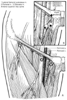 Figure 44-13
A, Anatomic landmarks
for lateral femoral cutaneous, femoral, and obturator nerve blocks. B,
For an obturator nerve block, the needle is walked off the inferior pubic ramus in
a medial and cephalad direction until it passes into the obturator canal (see Plate
12
in the color atlas of this volume).
Figure 44-13
A, Anatomic landmarks
for lateral femoral cutaneous, femoral, and obturator nerve blocks. B,
For an obturator nerve block, the needle is walked off the inferior pubic ramus in
a medial and cephalad direction until it passes into the obturator canal (see Plate
12
in the color atlas of this volume).
tubercle, identifying the inguinal ligament. The femoral artery is marked. A 22-gauge,
4-cm needle is advanced lateral to this line ( Fig.
44-13A
; see Plate 12
in the color atlas of this volume). When the needle reaches the depth of the artery,
a pulsation of the hub is visible. Elicitation of a paresthesia or motor response
verifies correct needle position. Commonly, the anterior branch of the femoral nerve
is identified first. Stimulation of this branch leads to contraction of the sartorius
muscle on the medial aspect of the thigh and should not be accepted. The needle
should be redirected slightly laterally and with a deeper direction to encounter
the posterior branch of the femoral nerve. Stimulation of this branch is identified
by patellar ascension as the quadriceps contract. Local anesthetic (20 mL) is injected
at that site.
Side Effects and Complications
Intravascular injection and hematoma are possible because of the
proximity of the femoral artery. Anatomically, the nerve and artery are located
in separate sheaths approximately 1 cm apart. In most patients with normal
anatomy, the femoral artery can be easily palpated, allowing correct, safe needle
positioning lateral to the pulsation. The presence of femoral vascular grafts is
a relative contraindication to this block. Nerve damage is rare with this technique.