LOWER EXTREMITY BLOCKS
Knowledge of the anatomy of the lumbosacral plexus and peripheral
nerves of the lower extremity enables anesthesiologists to provide more comprehensive
anesthetic care. These blocks are safe and have certain advantages, such as postoperative
pain relief and lack of complete sympathectomy, which make them ideal for selected
patients.
Lower extremity blocks are less popular than those routinely employed
for surgical procedures of the upper extremity. In part, this is because of the
widespread acceptance and safety of spinal and epidural anesthesia. Unlike the brachial
plexus, the nerves supplying the lower extremity are not anatomically clustered where
they can be easily blocked with a relatively superficial injection of local anesthetic.
Because of the anatomic considerations, lower extremity blocks are technically more
difficult and require more training and practice before expertise is acquired. Many
of these blocks were classically performed using paresthesia, loss of resistance,
or field block technique, and success rates varied. Advances in needles, catheters,
and nerve stimulator technology have facilitated localization of neural structures
and improved success rate. Recent applications have focused on prolonged postoperative
analgesia to assist rehabilitation and hospital dismissal.
Anatomy
The nerve supply to the lower extremity is derived from the lumbar
and sacral plexuses. The lumbar plexus is formed by the anterior rami of the first
four lumbar nerves, frequently including a branch from T12 and occasionally from
L5 ( Fig. 44-11
; see Plate
11
in the color atlas of this volume). The plexus lies between the psoas
major and quadratus lumborum muscles in the so-called psoas compartment.
The lower components of the plexus, L2, L3, and L4, primarily
innervate the anterior and medial thigh. The anterior divisions of L2, L3, and L4
form the obturator nerve; the posterior divisions of the same components form the
femoral nerve; and the lateral femoral cutaneous nerve is formed from posterior divisions
of L2 and L3.
The posterior cutaneous nerve of the thigh and the sciatic nerve
are derived from the first, second, and third sacral nerves plus branches from the
anterior rami of L4 and L5, respectively. These nerves pass together through the
pelvis and the greater sciatic foramen and are blocked by the same technique. The
sciatic nerve is a combination of two major nerve trunks, the tibial (i.e., ventral
branches
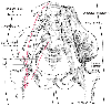 Figure 44-11
The lumbar plexus lies in the psoas compartment between
the psoas major and quadratus lumborum muscles (see Plate
11
in the color atlas of this volume).
Figure 44-11
The lumbar plexus lies in the psoas compartment between
the psoas major and quadratus lumborum muscles (see Plate
11
in the color atlas of this volume).
of the anterior rami of L4, L5, S1, S2, and S3) and the common peroneal (i.e., dorsal
branches of the anterior rami of L4, L5, S1, S2, and S3), which form the sciatic
nerve. At or above the popliteal fossa, they separate, with the tibial nerve passing
medially and the common peroneal laterally. The cutaneous distributions of the lumbosacral
and peripheral nerves are shown in Figure
44-12
.