|
|
|
|
|
|
|
|
|
|
|
|
|
|
|
The axillary approach to the brachial plexus is the most popular because of its ease, reliability, and safety.[20] Blockade occurs at the level of the terminal nerves. Although blockade of the musculocutaneous nerve is not
Anatomic concepts that should be considered before an axillary block include the following:
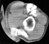 Figure 44-6
Axillary block. Computed tomogram after an axillary
block with 0.5% bupivacaine combined with iodothalamate. Separate 10-mL injections
of solution were made after obtaining median and radial nerve paresthesias transarterially.
Contrast medium appears to remain in three separate compartments.
Figure 44-6
Axillary block. Computed tomogram after an axillary
block with 0.5% bupivacaine combined with iodothalamate. Separate 10-mL injections
of solution were made after obtaining median and radial nerve paresthesias transarterially.
Contrast medium appears to remain in three separate compartments.
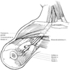 Figure 44-7
Axillary block. The arm is abducted at right angles
to the body. Distal digital pressure is maintained during needle placement and injection
of the local anesthetic (see Plate
5
in the color atlas of this volume).
Figure 44-7
Axillary block. The arm is abducted at right angles
to the body. Distal digital pressure is maintained during needle placement and injection
of the local anesthetic (see Plate
5
in the color atlas of this volume).
The patient should be in the supine position with the arm to be blocked placed at a right angle to the body and the elbow flexed to 90 degrees. The dorsum of the hand rests on the bed or pillow; hyperabduction of the arm with placement of the hand beneath the patient's head is not recommended because this position frequently obliterates the pulse.
The axillary artery is palpated, and a line is drawn tracing its course from the lower axilla as far proximally as possible. The artery is then fixed against the patient's humerus by the index and middle fingers of the left hand, and a skin wheal is raised directly over the artery at a point in the axilla approximating the skin crease. Proximal needle placement and maintenance of distal pressure facilitate proximal spread of the solution.
Several methods of identifying the axillary sheath have been described, all with reportedly good results. Overall, paresthesias are unnecessary. However, multiple injections may shorten the onset and may improve the reliability of blockade.
When the injection is completed, the arm should be adducted and returned to the patient's side. This prevents the humeral head from obstructing proximal flow of the solution; distal pressure and massage may also help. Vester-Andersen and colleagues[28] were unable to consistently block the musculocutaneous nerve with volumes up to 80 mL. If the musculocutaneous nerve is not blocked by the axillary approach, it can be blocked by injection within the body of the coracobrachialis muscle or at the elbow superficially at the lateral aspect of the antecubital fossa just above the interepicondylar line.
The success rate for an axillary block depends on the definition of a successful block (i.e., surgical anesthesia versus blockade of all four terminal nerves of the upper extremity), the technique used to localize the brachial plexus, and the number of injections. Success rates with single-injection techniques can vary.[29] [30] Thompson and Rorie[24] concluded that the presence of multiple compartments limits diffusion of the solution (and the success of single-shot techniques). Although Partridge and coworkers[31] confirmed the presence of these compartments, they concluded that the “septa” dividing them were incomplete on the basis of injections of methylene blue and latex solutions into cadavers. The controversy surrounding single- versus multiple-injection techniques remains unresolved.
Eliciting a paresthesia is as efficacious as peripheral nerve stimulation (with a motor response of 0.5 to 0.8 mA). Most studies suggest that two-injection transarterial techniques are equivalent to single-paresthesia or single-nerve stimulation approaches. In general, the efficacy of paresthesia and peripheral nerve stimulator techniques increases when multiple injections are employed. Conversely, success rates with perivascular or fascial click approaches are variously reliable. [7] Familiarity with a variety of techniques for axillary block of the brachial plexus allows the anesthesiologist maximal flexibility in tailoring the anesthetic approach to the clinical situation.
Nerve injury and systemic toxicity are the most significant complications associated with the axillary approach. The assertion that neuropathies are more common with the paresthesia technique may be valid, but it is not supported by the available data. Even when paresthesias are not sought, they often occur unintentionally. [32] Injection of large volumes of local anesthetic, particularly with the transarterial approach, increases the risk of intravascular injection and systemic toxicity of local anesthetics. Hematoma and infection are rare complications. Central neural blockade and pneumothorax are not complications, as in other approaches to the brachial plexus.
|
|
|
|
|
|
|
|
|
|
|
|
|