Temperature-Monitoring Sites
The core thermal compartment is composed of highly perfused tissues
whose temperature is uniform and high in comparison to the rest of the body. Temperature
in this compartment can be evaluated in the pulmonary artery, distal part of the
esophagus, tympanic membrane, or nasopharynx.[221]
[222]
Temperature probes incorporated into esophageal
stethoscopes must be positioned at the point of maximal heart sounds, or even more
distally, to provide accurate readings.[165]
Even
during rapid thermal perturbations (e.g., cardiopulmonary bypass), these temperature-monitoring
sites remain reliable. Core temperature can be estimated with reasonable accuracy
by using oral, axillary, rectal, and bladder temperatures, except during extreme
thermal perturbations.[221]
[222]
Skin surface temperatures are considerably lower than core temperature.
[223]
Skin surface temperatures—when adjusted
with an appropriate offset—nonetheless reflect core temperature reasonably
well.[224]
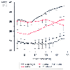
However, skin temperatures fail to reliably
confirm the clinical signs of malignant hyperthermia (tachycardia and hypercarbia)
in swine[225]
and have not been evaluated for this
purpose in humans ( Fig. 40-25
).
[225]
Rectal temperature also normally correlates
well with core temperature,[221]
[222]
but it fails to increase appropriately during malignant hyperthermia crises[225]
and in other documented situations.[226]
[227]
Consequently, rectal and skin surface temperatures must be used with some caution.
Even during cardiopulmonary bypass, the core temperature-monitoring
sites (e.g., tympanic membrane, nasopharynx, pulmonary artery, and esophagus) remain
useful. In contrast, rectal temperatures lag behind those measured in core sites.
Consequently, rectal temperature is considered an "intermediate" temperature in
deliberately cooled patients. During cardiac surgery, bladder temperature is equal
to rectal temperature (and therefore intermediate temperature) when urine flow is
low, but equal to pulmonary artery temperature (and thus core temperature) when flow
is high.[228]
Because bladder temperature is strongly
influenced by urine flow, it may be difficult to interpret in these patients. The
adequacy of
 Figure 40-25
Axillary and esophageal temperatures correlated well
during acute malignant hyperthermia in swine, but forehead and neck skin temperatures
did not. Rectal temperature also failed to promptly identify the onset of malignant
hyperthermia. Elapsed time zero indicates an end-tidal PCO2
of 70 mm Hg. These data indicate that forehead and neck skin surface temperatures
will not adequately confirm other clinical signs of malignant hyperthermia. Valid
core temperature monitoring sites include the distal part of the esophagus, pulmonary
artery, nasopharynx, and tympanic membrane. Except during cardio-pulmonary bypass,
body temperature can also be measured in the mouth, axilla, and bladder. Data are
presented as means ± SD. (Redrawn with modification from laizzo PA,
Zink RS, Kehler CH, et al: Skin and central temperature during malignant hyperthermia
in swine [abstract]. Anesthesiology 77:A569, 1992.)
rewarming is best evaluated by considering both "core" and "intermediate" temperatures.
 Figure 40-25
Axillary and esophageal temperatures correlated well
during acute malignant hyperthermia in swine, but forehead and neck skin temperatures
did not. Rectal temperature also failed to promptly identify the onset of malignant
hyperthermia. Elapsed time zero indicates an end-tidal PCO2
of 70 mm Hg. These data indicate that forehead and neck skin surface temperatures
will not adequately confirm other clinical signs of malignant hyperthermia. Valid
core temperature monitoring sites include the distal part of the esophagus, pulmonary
artery, nasopharynx, and tympanic membrane. Except during cardio-pulmonary bypass,
body temperature can also be measured in the mouth, axilla, and bladder. Data are
presented as means ± SD. (Redrawn with modification from laizzo PA,
Zink RS, Kehler CH, et al: Skin and central temperature during malignant hyperthermia
in swine [abstract]. Anesthesiology 77:A569, 1992.)