THE PROCESSED ELECTROENCEPHALOGRAM
Interpretation of the standard paper electroencephalographic tracing
is a science and an art. All monitored waveforms during the case are compared with
baseline signals. The interpreter has learned from experience the wide variety of
normal changes that may occur in the perioperative period and promptly recognizes
when changes occur that are not normal or expected. The baseline recordings and
the qualitative overall impression of the record are important in interpreting the
intraoperative EEG. Until recently, this qualitative approach was used because the
waveforms could not be described mathematically in a timeframe that would make such
information of any practical use. Analog-to-digital conversion technology associated
with mainframe computers was used to convert the analog signal of the EEG to digital
data and to mathematically manipulate the data. The process was complex, expensive,
and still had little relevance to the clinician. Early techniques took 1 hour to
digitize and analyze a 1-second epoch of electroencephalographic data. Computer
hardware has dramatically improved in speed and size, and real-time signal processing
is now possible and commonly used.
Several limitations are introduced when moving from the raw electroencephalographic
domain to the processed electroencephalographic domain. First, as the processed
electroencephalographic signal becomes more electronically remote (i.e., more processed),
there is a point at which it becomes increasingly difficult or impossible to relate
what we know about the raw electroencephalographic data to the processed signal.
An example of this problem was found in an early prototype electroencephalographic
monitor used in the operating room. When the dominant electroencephalographic frequency
and amplitude were kept in an acceptable reference range, a green light was visible.
When values fell outside this range, a red light appeared. Most of the valuable
information contained in the EEG was not visible to the clinician, and the displayed
lights were of little value in many cases. Today, some clinicians with no experience
in interpreting raw electroencephalographic data are using a processed EEG with little
ability to understand how it relates to the original raw data and how artifacts may
contaminate the signal and appear as perfectly believable processed electroencephalographic
data. Second, the standard 16-channel electroencephalographic montage provides more
information than can be practically analyzed or displayed in most processed electroencephalographic
monitors and perhaps more than is needed for routine intraoperative use. Studies
have not elucidated the optimal number of electroencephalographic channels for intraoperative
monitoring, but most available processed electroencephalographic devices use four
or fewer channels of information—translating to at most two channels per hemisphere.
Processed electroencephalographic devices generally monitor less cerebral territory
than a standard 16-channel EEG. Third, some intraoperative changes are unilateral,
and some are bilateral. Display of the activity of both hemispheres is necessary
to differentiate unilateral (i.e., not caused by global factors such as anesthesia)
from bilateral changes. An appropriate number of leads over both hemispheres is
needed. The gold standard for intraoperative electroencephalographic monitoring
is the continuous visual inspection of a 16- to 32-channel analog EEG by an experienced
electroencephalographer.[10]
[11]
Adequate studies comparing processed EEG with fewer channels to this gold standard
across multiple uses and operations have not been done, although limited data using
processed electroencephalographic monitoring during carotid surgery suggest that
two- or four-channel instruments can detect most significant changes.[12]
[13]
Devices
Two basic forms of electroencephalographic processing are used:
power analysis and bispectral analysis. Power analysis uses Fourier transformation
to convert the digitized raw electroencephalographic signal into component sine waves
of identifiable frequency and amplitude. The raw electroencephalographic data, which
are plots of voltage versus time, are converted to plots of frequency and amplitude
versus time. Many commercially available processed electroencephalographic machines
display power (i.e., voltage or amplitude squared) as a function of frequency and
time. These monitors display the data in two general forms: compressed spectral
array (CSA) or density spectral array (DSA). In CSA, frequency is displayed along
the x axis, and power is displayed along the y
axis, with the height of the waveform equal to the
power at that frequency. Time is displayed along the z
axis. Tracings overlap each other, with the most recent information in front ( Fig.
38-4
). DSA also displays frequency along the x
axis and time along the y axis, and power is reflected
by the density of the dots at each frequency. Each display format provides the same
data, and the choice depends on the preference of the user.
Many changes that occur during anesthesia and surgery are reflected
as changes in amplitude or frequency, or both. These changes can be clearly seen
in the displays if adequate and appropriate channels are monitored. Power analysis
has been used clinically for many years as a diagnostic tool during procedures with
a risk for intraoperative cerebral ischemia such as carotid endarterectomy and cardiopulmonary
bypass (CPB). Power analysis has proved to be a sensitive and reliable monitor in
the hands of experienced operators using an adequate number of channels. Parameters
obtained from power analysis have been investigated as monitors for depth of anesthesia.
[14]
[15]
[16]
[17]
Although earlier attempts to use parameters
derived from power analysis for assessment of anesthetic depth were largely unsuccessful,
these same parameters are now used to various degrees with much more success as a
part of different algorithms (including BIS) to measure hypnotic states.
Bispectral analysis takes into account the phase relationships
between the individual components of the raw electroencephalographic signal. These
phase relationships
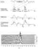 Figure 38-4
Diagram of the technique used to generate a compressed
spectral array. Below the traces, the example shows compressed spectra of the α
rhythm from a normal subject. (From Stockard JJ, Bickford RG: The neurophysiology
of anesthesia. In Gordon E [ed]: A Basis and Practice
of Neuroanesthesia. New York, Elsevier, 1981, p 3.)
Figure 38-4
Diagram of the technique used to generate a compressed
spectral array. Below the traces, the example shows compressed spectra of the α
rhythm from a normal subject. (From Stockard JJ, Bickford RG: The neurophysiology
of anesthesia. In Gordon E [ed]: A Basis and Practice
of Neuroanesthesia. New York, Elsevier, 1981, p 3.)
are not included in power analysis. Bispectral analysis has seen extensive use in
the past decade, and the primary use of this analysis technique is as a monitor of
depth of hypnosis. Although the bispectral analysis may also yield information suggestive
of cerebral pathologic states developing intraoperatively, such as cerebral ischemia,
it is not recommended by the manufacturer for this use.[18]
[19]
[20]