Intraoperative or Procedural Management of Implantable
Cardioverter-Defibrillators
No special monitoring is required for the patient with an ICD.
Electrocardiographic monitoring and the ability to deliver external cardioversion
or defibrillation must be present during the time of ICD disablement. Should external
cardioversion or defibrillation be needed, the defibrillator pads should be placed
to avoid the pulse generator and lead system to the extent possible. Nevertheless,
one should remember that the patient, not the ICD, is being treated. The recommendations
in the section "Intraoperative or Procedural Management of Pacemakers" apply here
as well.
No special anesthetic techniques have been championed for the
patient with an ICD. Most of these patients will have severely depressed systolic
function, dilated ventricular cavities, and significant valvular regurgitation.
The choice of anesthetic technique should be dictated by the underlying physiologic
derangements that are present. Conflicting data have been published regarding the
choice of anesthetic agents and changes to defibrillation threshold (DFT). In 1992,
Gill and colleagues examined DFT in dogs and concluded that neither halothane nor
isoflurane changed DFT in open chest defibrillation compared to a pentobarbital infusion.
[87]
However, Weinbroum and associates recently
evaluated defibrillation thresholds in humans during ICD implant
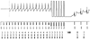 Figure 35-8
Unexpected ventricular tachycardia with antitachycardia
pacing (ATP) was found in this patient during her preoperative visit. A 65-year-old
woman with a history of ventricular tachycardia (VT) had undergone implantation of
a Medtronic single-chamber defibrillator about 8 months earlier. She had no dizziness
or syncopal episodes since the implantable cardioverter-defibrillator (ICD) placement.
Interrogation of her device in the preoperative center revealed VVE-VVI programming,
along with an episode of tachycardia at 150 to 162 beats/min that was detected by
the ICD as VT. The ICD delivered a 6-beat burst of antitachycardia pacing at 182
beats/min, which converted the tachycardia back to sinus rhythm. No backup antibradycardia
pacing was needed after the VT was terminated. The upper tracing
is a digitized ventricular electrogram that was stored in the ICD during the tachycardic
event. The lower tracing is the marker channel that
reports the interpretation of the ICD for each event. The numbers below the marker
channel represent the interval (in milliseconds). The heart rate is calculated by
dividing the interval into 60,000 msec/min. TS represents an interval in the VT
zone, TD marks the final event that starts therapy, TP is an ATP event, and VS is
an intrinsic ventricular depolarization with a rate that is neither too fast (short
interval) nor too slow (long interval). This device was set to detect VT as 16 consecutive
ventricular events with a rate between 146 and 200 beats/min and to deliver ATP at
84% of the last R-R interval. The last interval was 400 msec, so ATP was delivered
at a rate of 182 beats/min (330-msec intervals).
Figure 35-8
Unexpected ventricular tachycardia with antitachycardia
pacing (ATP) was found in this patient during her preoperative visit. A 65-year-old
woman with a history of ventricular tachycardia (VT) had undergone implantation of
a Medtronic single-chamber defibrillator about 8 months earlier. She had no dizziness
or syncopal episodes since the implantable cardioverter-defibrillator (ICD) placement.
Interrogation of her device in the preoperative center revealed VVE-VVI programming,
along with an episode of tachycardia at 150 to 162 beats/min that was detected by
the ICD as VT. The ICD delivered a 6-beat burst of antitachycardia pacing at 182
beats/min, which converted the tachycardia back to sinus rhythm. No backup antibradycardia
pacing was needed after the VT was terminated. The upper tracing
is a digitized ventricular electrogram that was stored in the ICD during the tachycardic
event. The lower tracing is the marker channel that
reports the interpretation of the ICD for each event. The numbers below the marker
channel represent the interval (in milliseconds). The heart rate is calculated by
dividing the interval into 60,000 msec/min. TS represents an interval in the VT
zone, TD marks the final event that starts therapy, TP is an ATP event, and VS is
an intrinsic ventricular depolarization with a rate that is neither too fast (short
interval) nor too slow (long interval). This device was set to detect VT as 16 consecutive
ventricular events with a rate between 146 and 200 beats/min and to deliver ATP at
84% of the last R-R interval. The last interval was 400 msec, so ATP was delivered
at a rate of 182 beats/min (330-msec intervals).
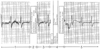 Figure 35-9
Electromagnetic interference from the monopolar electrosurgery
(i.e., Bovie) caused an implantable cardioverter-defibrillator (ICD) to detect ventricular
fibrillation (VF). This stored electrogram was one of 73 found at the end of a 4-hour
surgical procedure in which considerable monopolar electrosurgery (ESU) was used.
This patient had a Guidant Medical ICD in the VOE-VVI mode. This patient's ICD
had been placed in a "monitor only" mode before surgery. As a result, the ICD recorded
any instance of ventricular dysrhythmia that would have triggered therapy, but it
could not deliver therapy. The electrogram demonstrates a ventricular rate of 70
beats/min but with considerable noise on the baseline (1); a ventricular fibrillation
(VF) event was declared for the detected heart rate of 345 beats/min, and the ICD
charged its capacitor (2); the ICD was programmed to "reconfirm before shock," and
the ventricular rate remains 70 beats/min with noise on the baseline (3); the noise
caused the ICD to believe that the patient remained in VF, and the ICD would have
delivered a shock, except it was programmed to monitor only (4); and because the
noise is gone (i.e., the ESU had stopped), the ICD declares the event over after
a "successful" defibrillation (5).
Figure 35-9
Electromagnetic interference from the monopolar electrosurgery
(i.e., Bovie) caused an implantable cardioverter-defibrillator (ICD) to detect ventricular
fibrillation (VF). This stored electrogram was one of 73 found at the end of a 4-hour
surgical procedure in which considerable monopolar electrosurgery (ESU) was used.
This patient had a Guidant Medical ICD in the VOE-VVI mode. This patient's ICD
had been placed in a "monitor only" mode before surgery. As a result, the ICD recorded
any instance of ventricular dysrhythmia that would have triggered therapy, but it
could not deliver therapy. The electrogram demonstrates a ventricular rate of 70
beats/min but with considerable noise on the baseline (1); a ventricular fibrillation
(VF) event was declared for the detected heart rate of 345 beats/min, and the ICD
charged its capacitor (2); the ICD was programmed to "reconfirm before shock," and
the ventricular rate remains 70 beats/min with noise on the baseline (3); the noise
caused the ICD to believe that the patient remained in VF, and the ICD would have
delivered a shock, except it was programmed to monitor only (4); and because the
noise is gone (i.e., the ESU had stopped), the ICD declares the event over after
a "successful" defibrillation (5).
and found that halothane, isoflurane, and fentanyl increased DFT.[88]
Even with these increases, the increased DFTs found were still substantially lower
than the maximum energy generally available in ICDs, and these increases would not
have been noted under usual testing conditions.